Review
. 2010 Apr;120(4):963-72.
doi: 10.1172/JCI41350. Epub 2010 Apr 1.

The ovary: basic biology and clinical implications

Affiliations
Review

The ovary: basic biology and clinical implications

Joanne S Richards et al. J Clin Invest. 2010 Apr.

Abstract

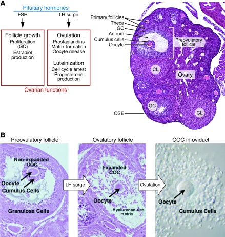
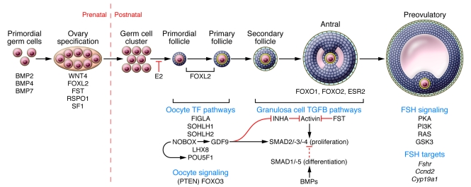
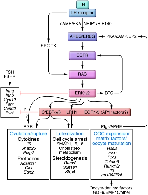
The classical view of ovarian follicle development is that it is regulated by the hypothalamic-pituitary-ovarian axis, in which gonadotropin-releasing hormone (GnRH) controls the release of the gonadotropic hormones follicle-stimulating hormone (FSH) and luteinizing hormone (LH), and that ovarian steroids exert both negative and positive regulatory effects on GnRH secretion. More recent studies in mice and humans indicate that many other intra-ovarian signaling cascades affect follicular development and gonadotropin action in a stage- and context-specific manner. As we discuss here, mutant mouse models and clinical evidence indicate that some of the most powerful intra-ovarian regulators of follicular development include the TGF-beta/SMAD, WNT/FZD/beta-catenin, and RAS/ERK1/2 signaling pathways and the FOXO/FOXL2 transcription factors.


Figures

Figure 1
Figure 1. Summary of hormonal control of the ovary during follicle growth, ovulation, and luteinization.
(A) Left: The pituitary gonadotropins FSH and LH are part of the hypothalamic-pituitary-gonadal axis that coordinately regulates the menstrual (in humans) or estrous (in nonhuman mammals) cycle by extensive feedback loops. FSH controls follicular granulosa cell (GC) growth and estradiol production, while LH controls ovulation and follicular luteinization. Right: A cross section of a mouse ovary is shown, demonstrating the main cell types and follicle stages. Ovarian follicles are composed of a single oocyte surrounded by somatic cells (granulosa cells) and thecal cells. Follicles grow from primordial (not shown) to primary and secondary stages independent of the pituitary gonadotropins. FSH stimulates growth to the preovulatory follicle stage, characterized by granulosa cells that directly surround the oocyte (cumulus cells) and those that make up the bulk of the wall of the follicle. Following the LH surge, the follicle erupts through the ovarian surface (OSE), and the remaining cells of the follicle terminally differentiate to form a corpus luteum. Original magnification, ×5. (B) Ovulation in the mouse. Left: The preovulatory follicle contains an oocyte surrounded by cumulus cells that are separated from the mural granulosa cells by a fluid-filled cavity. Middle: Following the LH surge, the COC undergoes a process called cumulus or COC expansion, in which the cumulus cells make and become embedded in a hyaluronan-rich matrix. Right: The cumulus cells accompany the oocyte into the oviduct following release of the entire COC from the ovarian follicle.Original magnification, ×40.
Figure 2
Figure 2. Signaling pathways controlling ovarian follicle growth in the mouse.
PGCs are specified by the BMP pathway, then proliferate and migrate to the indifferent gonad. The BMPs are major determinants of PGC specification and proliferation in the mouse. During the postnatal period, clusters (or nests) of germ cells break down to form primordial follicles, which upon activation become primary follicles. Estradiol (E2) inhibits the breakdown of germ cell clusters to primordial follicles. A number of mice lacking oocyte transcription factors (TFs) (NOBOX, SOHLH1, SOHLH2, and LHX8) show loss of follicles at the primordial follicle–to–primary follicle transition or before primordial follicle formation (FIGLA). Foxo3 is also expressed in oocytes, and deletion of Foxo3 (or the PI3K inhibitor Pten) in oocytes results in premature activation of the primordial follicle pool and oocyte loss. Foxl2 is expressed in somatic granulosa cells, and deletion results in arrest and subsequent death of follicles before the primary follicle stage. FOXL2 also likely functions at late stages in folliculogenesis. NOBOX regulates other TFs (e.g., Pou5f1) and also Gdf9, the product of which is secreted by the oocyte to regulate granulosa cell function, including suppression of Inha. GDF9 and activin signal though SMAD2/-3. Regulation of activin by its inhibitors, such as inhibin or follistatin, is critical after the secondary follicle stage and involves many follicle stages. In vitro experiments implicate FOXO1 and ESR2 as important regulators in granulosa cells of growing follicles. FSH is a key regulator of the antral and preovulatory stage through multiple signaling cascades, and many of the FSH targets at the preovulatory stage are coregulated by activin (Fshr, Cyp19a1) and/or β-catenin (Cyp19a1). See text for discussion and references for each indicated gene pathway.
Figure 3
Figure 3. LH-mediated pathways to ovulation and luteinization.
LH induces ovulation, COC expansion, oocyte maturation, and luteinization in preovulatory follicles. These events are mediated by LH activation of the PKA pathway and NRIP1, which induce the expression of the EGF-like factors (AREG, EREG). AREG and/or EREG in turn activate the EGFR signaling cascade, including RAS and ERK1/2. This hypothesis is based on the observations that when Erk1 and Erk2 are disrupted in granulosa cells, global changes occur in gene expression patterns that control ovulation, COC expansion, resumption of meiosis, and luteinization. Moreover, activation of ERK1/2 is essential to turn off the FSH-regulated gene expression program that controls genes essential for preovulatory follicle growth and differentiation. C/EBPα and C/EBPβ, NR5A2 (also known as LRH1), and possibly PGR appear to be among the key transcription factors that are activated by ERK1/2 phosphorylation and affect ovulation and luteinization. Other transcription factors that are targets of ERK1/2 include members of the AP1 family and EGR1/3, whose specific functions in ovulation and luteinization remain to be defined. Additionally, oocyte-derived factors such as GDF9 and BMP15 affect cumulus cell and granulosa cell functions in a gradient-dependent manner. The functions of specific genes are discussed in the text. TK, tyrosine kinase.

Similar articles

Cited by

References

    1. De Felici M, et al. Experimental approaches to the study of primordial germ cell lineage and proliferation. Hum Reprod Update. 2004;10(3):197–206. doi: 10.1093/humupd/dmh020. - DOI - PubMed
    1. Pesce M, Gioia Klinger F, De Felici M. Derivation in culture of primordial germ cells from cells of the mouse epiblast: phenotypic induction and growth control by Bmp4 signalling. Mech Dev. 2002;112(1-2):15–24. doi: 10.1016/S0925-4773(01)00624-4. - DOI - PubMed
    1. Puglisi R, Montanari M, Chiarella P, Stefanini M, Boitani C. Regulatory role of BMP2 and BMP7 in spermatogonia and Sertoli cell proliferation in the immature mouse. Eur J Endocrinol. 2004;151(4):511–520. doi: 10.1530/eje.0.1510511. - DOI - PubMed
    1. Ross A, Munger S, Capel B. Bmp7 regulates germ cell proliferation in mouse fetal gonads. Sex Dev. 2007;1(2):127–137. doi: 10.1159/000100034. - DOI - PubMed
    1. Martins da Silva SJ, Bayne RA, Cambray N, Hartley PS, McNeilly AS, Anderson RA. Expression of activin subunits and receptors in the developing human ovary: activin A promotes germ cell survival and proliferation before primordial follicle formation. Dev Biol. 2004;266(2):334–345. doi: 10.1016/j.ydbio.2003.10.030. - DOI - PubMed

Publication types

Substances