

Review
doi: 10.1172/JCI41585.
Epub 2010 Apr 1.
Fertilization: a sperm's journey to and interaction with the oocyte
Affiliations
- PMID: 20364096
- PMCID: PMC2846064
- DOI: 10.1172/JCI41585
Item in Clipboard
Review
Fertilization: a sperm's journey to and interaction with the oocyte
J Clin Invest.
2010 Apr.
Abstract
Mammalian fertilization comprises sperm migration through the female reproductive tract, biochemical and morphological changes to sperm, and sperm-egg interaction in the oviduct. Recent gene knockout approaches in mice have revealed that many factors previously considered important for fertilization are largely dispensable, or if they are essential, they have an unexpected function. These results indicate that what has been observed in in vitro fertilization (IVF) differs significantly from what occurs during "physiological" fertilization. This Review focuses on the advantages of studying fertilization using gene-manipulated animals and highlights an emerging molecular mechanism of mammalian fertilization.
Figures
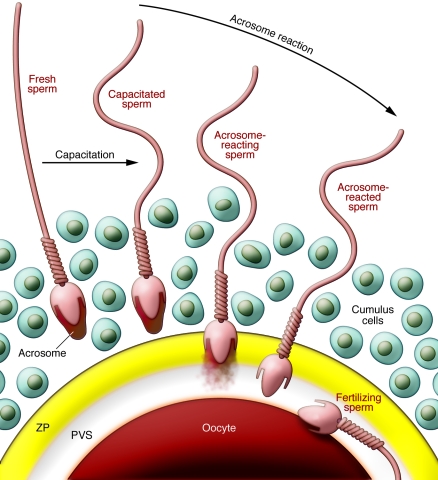
Over the nucleus of each mammalian sperm is a membranous sac known as the acrosome, which is filled with many kinds of hydrolytic enzymes. In the female reproductive tract or in an IVF medium, sperm undergo capacitation, which permits the acrosome reaction. Near the eggs, probably stimulated by the cumulus cells and the ZP, sperm release their acrosomal contents by exocytosis and penetrate the ZP. Only acrosome-reacted sperm fuse with eggs, but their competency for fusion does not last long. Cumulus cells are packed together by hyaluronic acid at ovulation and become diffuse during fertilization. PVS, perivitelline space.
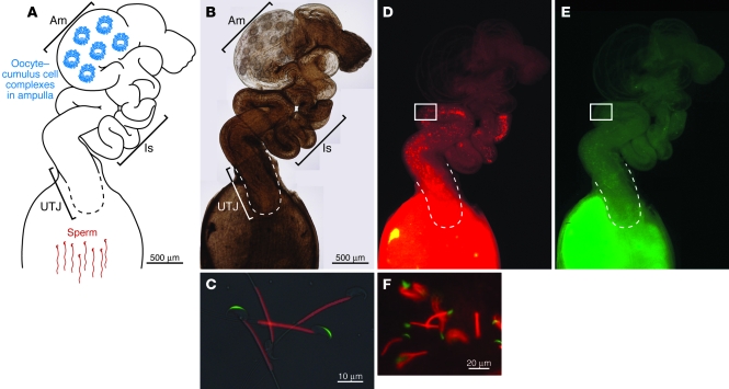
After sperm are deposited in the female reproductive environment, they become metabolically active and start migrating into the oviduct. (A and B) Depicted here is one oviduct in a female mouse. The generation of sperm engineered to express fluorescent proteins has facilitated visualization of sperm migration through the female reproductive tract (65, 66). (C) Sperm from mice engineered to express acrosome-targeted EGFP (acrEGFP) and mitochondria-targeted red fluorescent protein (mtDsRed2). GFP is released during the acrosome reaction, so that acrosome-reacted sperm are no longer acrEGFP+ but remain mtDsRed2+ (66). The mice are available through public bioresource centers. (B and D–F) The female mouse reproductive tract removed at four hours after coitus. The sperm and their acrosomal status can be monitored through the uterine and oviduct wall. Areas indicated in D and E are merged in F. The images in B, D, and E are a composite of several images. Scale bar: 500 μm (B, D, and E). Am, ampulla; Is, isthmus.
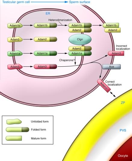
Disruption of the genes Clgn, Ace, Adam1a, Adam2, and Adam3 results in impaired sperm-ZP binding and impaired migration through the UTJ. Clgn is required for Adam1a/Adam2 and Adam1b/Adam2 heterodimerization. Lack of Adam1a/Adam2 heterodimerization in Clgn–/–, Adam1a–/–, and Adam2–/– mice causes Adam3 disappearance from the surface of mature sperm. Disruption of Ace leads to aberrant localization of Adam3, as evidenced by reduced amounts of Adam3 protein in the Triton X-114 detergent-enriched phase of sperm membranes (28). The diagram illustrates why disruption of the individual Ace, Clgn, Adam1a, Adam2, and Adam3 genes produces similar phenotypes and indicates the importance of Adam3 in sperm fertilizing ability.
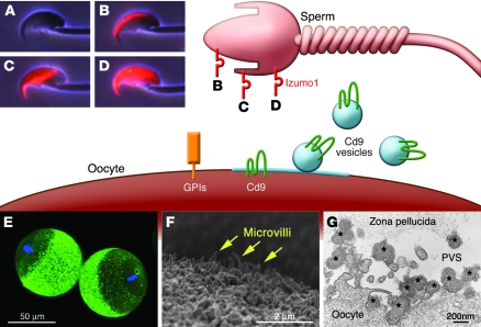
Sperm Izumo1 and egg Cd9 are essential factors for sperm-egg fusion (13, 16). GPI-anchored proteins on the egg surface are also essential (114), but none of the individual proteins have been identified. (A) Izumo1 is an acrosomal membrane protein that is not exposed before the acrosome reaction is complete. Acrosome-reacted sperm can be classified into three major groups by their Izumo1 staining pattern: acrosomal cap (B), equatorial (C), and whole head (D). (E and F) Cd9 is localized on the cilia distributed across the surface of unfertilized eggs, except the cilia at the metaphase plate. (G) Cd9-containing vesicles (asterisks) secreted from the oocyte are able to translocate onto the sperm surface and may play a role in sperm-egg fusion. G is reproduced with permission from Proceedings of the National Academy of Sciences of the United States of America (115).
References
-
- Chang MC. Fertilizing capacity of spermatozoa deposited into the fallopian tubes. Nature. 1951;168(4277):697–698. - PubMed
-
- Austin CR. Observations on the penetration of the sperm in the mammalian egg. Aust J Sci Res B. 1951;4(4):581–596. - PubMed
-
- Yanagimachi R, Chang MC. Fertilization of hamster eggs in vitro. Nature. 1963;200:281–282. - PubMed
Publication types
MeSH terms
Substances
LinkOut - more resources
Full Text Sources
Other Literature Sources
Molecular Biology Databases
