Review
. 2010 Apr;120(4):995-1003.
doi: 10.1172/JCI41229. Epub 2010 Apr 1.

Making the blastocyst: lessons from the mouse

Affiliations
Review

Making the blastocyst: lessons from the mouse

Katie Cockburn et al. J Clin Invest. 2010 Apr.

Abstract

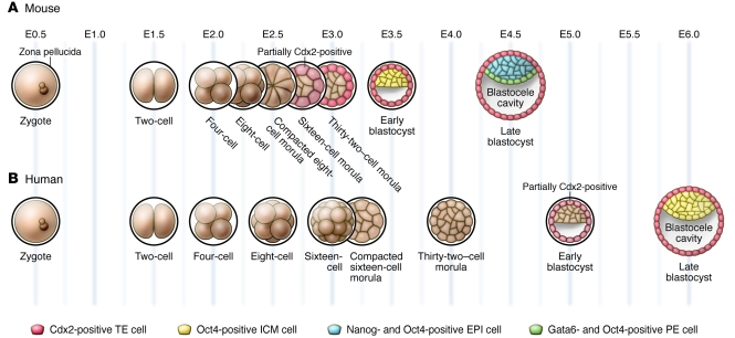
Mammalian preimplantation development, which is the period extending from fertilization to implantation, results in the formation of a blastocyst with three distinct cell lineages. Only one of these lineages, the epiblast, contributes to the embryo itself, while the other two lineages, the trophectoderm and the primitive endoderm, become extra-embryonic tissues. Significant gains have been made in our understanding of the major events of mouse preimplantation development, and recent discoveries have shed new light on the establishment of the three blastocyst lineages. What is less clear, however, is how closely human preimplantation development mimics that in the mouse. A greater understanding of the similarities and differences between mouse and human preimplantation development has implications for improving assisted reproductive technologies and for deriving human embryonic stem cells.


Figures

Figure 1
Figure 1. Stages of mouse and human preimplantation development.
(A) In the mouse, the fertilized egg undergoes three rounds of cleavage, producing an eight-cell embryo that then undergoes compaction. From the eight-cell stage onward, cell divisions produce two populations of cells, those that occupy the inside of the embryo and those that are located on the outside. The blastocoel cavity begins to form inside the embryo beginning at the 32-cell stage and continues to expand as the embryo grows and matures into the late blastocyst stage. Cdx2 becomes upregulated in outside, future TE cells, starting at the 32-cell stage, while Oct4 expression becomes limited to the ICM in the early blastocyst stage. By the late blastocyst stage, while continuing to express Oct4 ubiquitously, the ICM contains a population of Nanog-positive EPI cells and a population of Gata6-positive PE cells. (B) Development is similar in the early human embryo, although compaction occurs at the 16-cell stage and the mutually exclusive expression patterns of CDX2 and OCT4 are not established until the late blastocyst stage. The expression patterns of NANOG and GATA6 in the human preimplantation embryo have not yet been characterized.
Figure 2
Figure 2. Polarity in the mouse preimplantation embryo.
(A) At the eight-cell stage, all blastomeres polarize along the axis of cell contact, forming outward, apical domains and inward-facing basolateral domains. (B) As the embryo grows from eight to 16 cells, blastomeres that divide parallel to the inside-outside axis produce two outside, polar cells. Blastomeres that divide perpendicular to the inside-outside axis produce one outside, polar daughter cell and one non-polar, inside daughter cell. This creates two populations of cells: outside, polar cells and inside, nonpolar cells. These two types of cell division also occur as the embryo grows from 16 to 32 cells.
Figure 3
Figure 3. Models of TE specification in the mouse embryo.
(A) According to the inside-outside model, cells on the inside and outside of the embryo receive different amounts of cell contact, and this is translated into differences in transcription factor expression. (B) According to the cell polarity model, the presence or absence of an apical domain is translated into differences in transcription factor expression. (C) After the eight-cell stage, active Lats1/2 kinases phosphorylate Yap in inside cells, preventing its movement into the nucleus. Without Yap, Tead4 cannot induce the expression of Cdx2. In outside cells, Lat1 and Lat2 are inactive and Yap is free to move into the nucleus, activating Cdx2. Increased cell-cell contact on the inside of the embryo may activate Lat1 and Lat2 via the Hippo signaling pathway, while some component of the apical domain may inhibit Hippo signaling and Lat1 and Lat2 activity in outside cells.
Figure 4
Figure 4. Models of EPI/PE segregation in the mouse embryo.
(A) In the position-dependent model, the mouse ICM at E3.5 is composed of a uniform population of bipotential cells, and those cells located on the outside surface of the ICM become PE due to some form of positional information. (B) In the Fgf/MAPK-dependent model, cells of the ICM are initially bipotential, but differences in Fgf signaling cause them to become either Nanog- or Gata6-positive by E3.5. These cells are distributed randomly in the ICM, and cell sorting combined with apoptosis results in the formation of organized PE and EPI layers by E4.5.

Similar articles

Cited by

References

    1. Bacharova R. Gene expression during oogenesis and oocyte development in mammals. Dev Biol (N Y 1985). 1985;1:453–524. - PubMed
    1. Aoki F, Worrad DM, Schultz RM. Regulation of transcriptional activity during the first and second cell cycles in the preimplantation mouse embryo. Dev Biol. 1997;181(2):296–307. doi: 10.1006/dbio.1996.8466. - DOI - PubMed
    1. Hamatani T, Carter MG, Sharov AA, Ko MS. Dynamics of global gene expression changes during mouse preimplantation development. Dev Cell. 2004;6(1):117–131. doi: 10.1016/S1534-5807(03)00373-3. - DOI - PubMed
    1. Bachvarova R, Moy K. Autoradiographic studies on the distribution of labeled maternal RNA in early mouse embryos. J Exp Zool. 1985;233(3):397–403. doi: 10.1002/jez.1402330308. - DOI - PubMed
    1. Paynton BV, Rempel R, Bachvarova R. Changes in state of adenylation and time course of degradation of maternal mRNAs during oocyte maturation and early embryonic development in the mouse. Dev Biol. 1988;129(2):304–314. doi: 10.1016/0012-1606(88)90377-6. - DOI - PubMed

Publication types

Substances