

Review
doi: 10.1172/JCI41210.
Epub 2010 Apr 1.
Uterine disorders and pregnancy complications: insights from mouse models
Affiliations
- PMID: 20364098
- PMCID: PMC2846054
- DOI: 10.1172/JCI41210
Item in Clipboard
Review
Uterine disorders and pregnancy complications: insights from mouse models
J Clin Invest.
2010 Apr.
Abstract
Much of our knowledge of human uterine physiology and pathology has been extrapolated from the study of diverse animal models, as there is no ideal system for studying human uterine biology in vitro. Although it remains debatable whether mouse models are the most suitable system for investigating human uterine function(s), gene-manipulated mice are considered by many the most useful tool for mechanistic analysis, and numerous studies have identified many similarities in female reproduction between the two species. This Review brings together information from studies using animal models, in particular mouse models, that shed light on normal and pathologic aspects of uterine biology and pregnancy complications.
Figures

The uterus undergoes a sequence of cellular transformations every 28 days. Prior to ovulation, the uterus is at the follicular phase and has a thinner endometrium. Accompanied by peaks of follicle-stimulating hormone and estrogen, ovulation occurs mid-cycle. The uterine lining then proliferates and becomes predecidual for the preparation of blastocyst implantation. In humans, the receptive phase is considered to be around mid-luteal phase, at days 20–24 of each cycle (numbered in red).
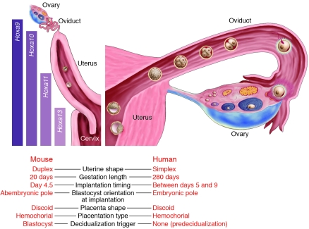
Columns on the left show distinct boundaries of Hoxa gene expression in the reproductive tract. Demarcation of expression boundaries is based on observations in the mouse; although HOXA genes are also implicated in human reproduction, whether the expression patterns are similar to those in mice is not known.

Schematic diagrams depicting cross-sections of implantation sites on days 4, 7, and 13 of pregnancy. (A) On day 4, the luminal epithelium closes on an implanting blastocyst. The mural trophectoderm, which is distant from the inner cell mass, contacts the epithelium at the antimesometrial side (AM). The decidual response also starts from the antimesometrial side (not shown). M, mesometrial side. (B) On day 7, the embryo is much larger, and its ectoplacental cone has penetrated the mesometrial decidua, which is enriched with blood vessels. Differentiated decidual cells now take up most of the implantation site (IS). Note that decidualization does not occur at inter-implantation sites. (C) On day 13, the placenta has developed, and the decidua has regressed to thin layers around the placenta and embryo, known as decidua basalis and decidua capsularis, respectively.
References
-
- Volpe EP. Developmental biology and human concerns. American Zoologist. 1987;27:697–714.
Publication types
MeSH terms
LinkOut - more resources
Full Text Sources
Other Literature Sources
Medical
Molecular Biology Databases
