. 2010 May;6(4):462-72.
doi: 10.4161/auto.6.4.11553. Epub 2010 May 16.

Cyclophilin D is required for mitochondrial removal by autophagy in cardiac cells

Affiliations

Cyclophilin D is required for mitochondrial removal by autophagy in cardiac cells

Raquel S Carreira et al. Autophagy. 2010 May.

Abstract

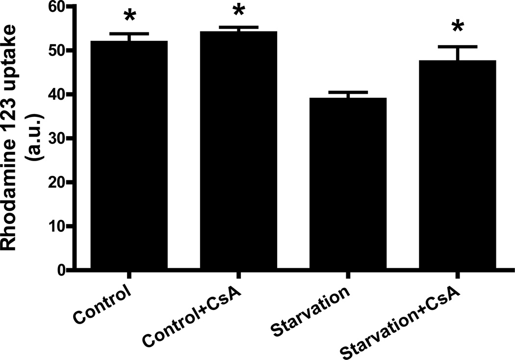
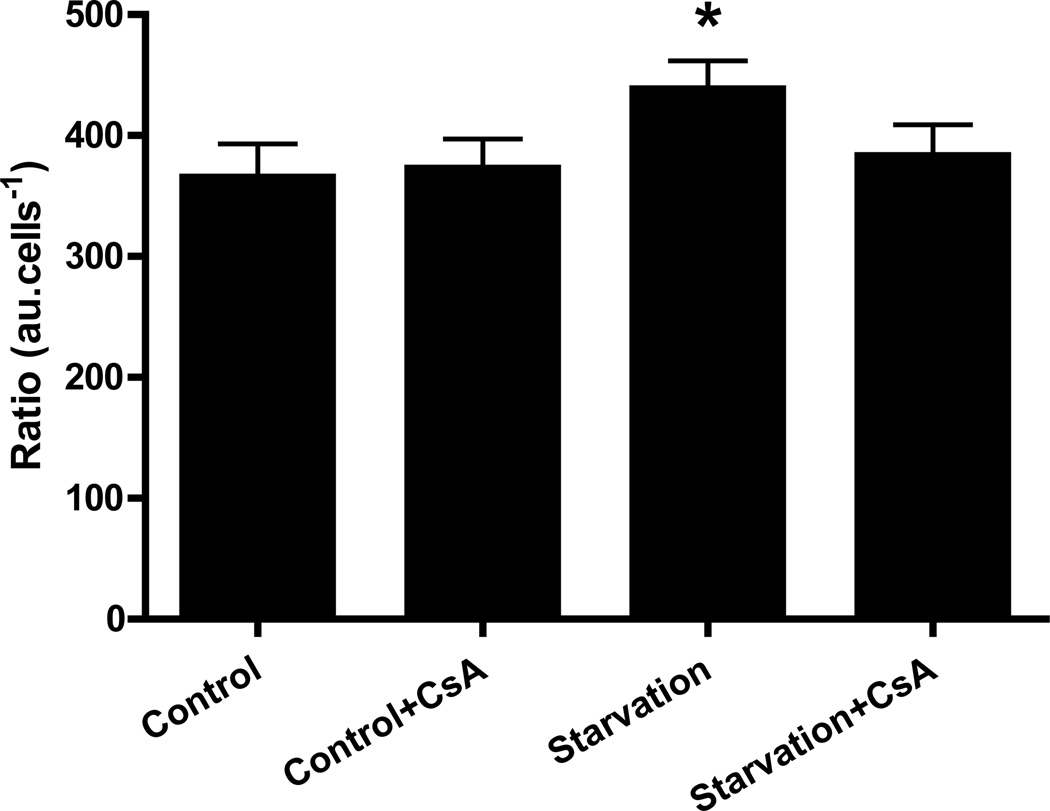
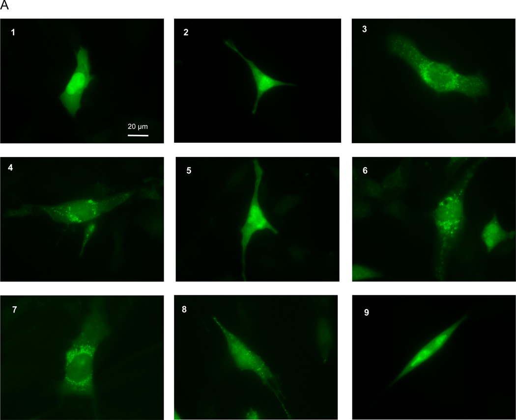
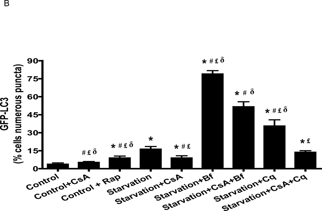
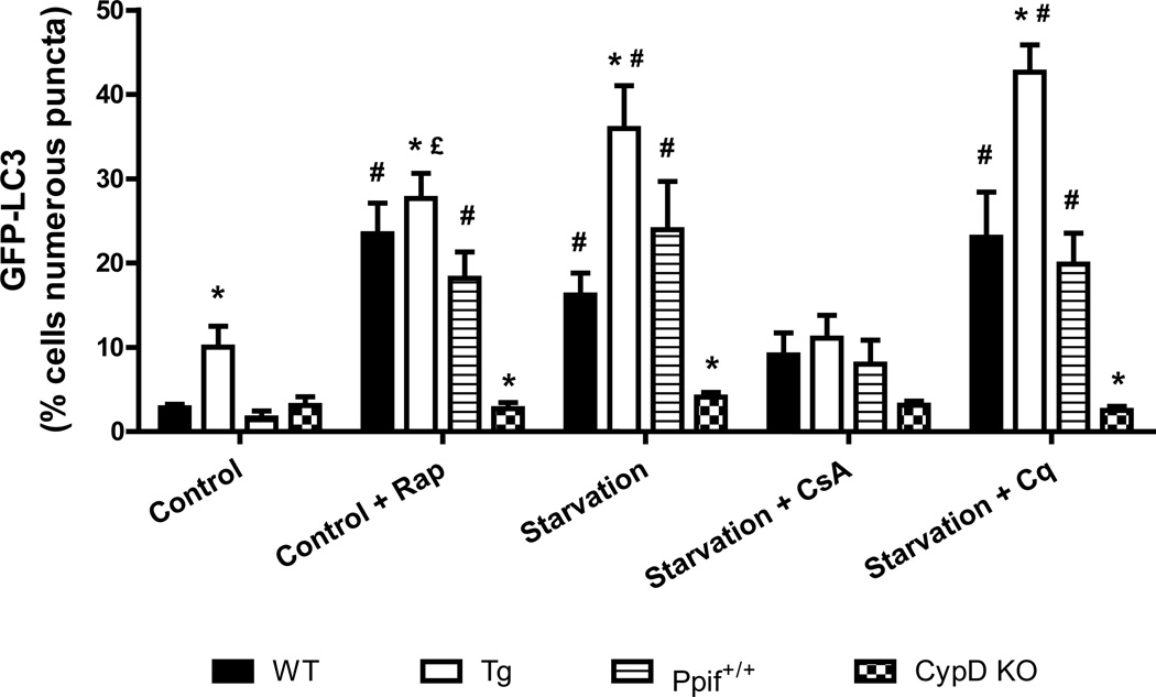
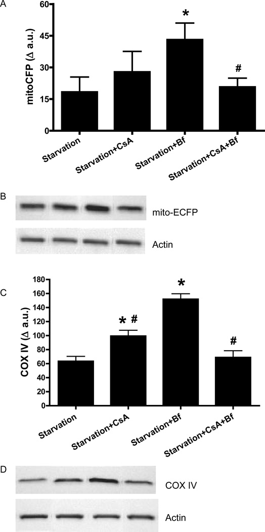
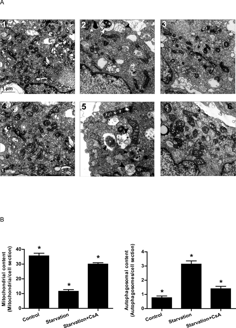
Autophagy is a highly regulated intracellular degradation process by which cells remove cytosolic long-lived proteins and damaged organelles. The mitochondrial permeability transition (MPT) results in mitochondrial depolarization and increased reactive oxygen species production, which can trigger autophagy. Therefore, we hypothesized that the MPT may have a role in signaling autophagy in cardiac cells. Mitochondrial membrane potential was lower in HL-1 cells subjected to starvation compared to cells maintained in full medium. Mitochondrial membrane potential was preserved in starved cells treated with cyclosporin A (CsA), suggesting the MPT pore is associated with starvation-induced depolarization. Starvation-induced autophagy in HL-1 cells, neonatal rat cardiomyocytes and adult mouse cardiomyocytes was inhibited by CsA. Starvation failed to induce autophagy in CypD-deficient murine cardiomyocytes, whereas in myocytes from mice overexpressing CypD the levels of autophagy were enhanced even under fed conditions. Collectively, these results demonstrate a role for CypD and the MPT in the initiation of autophagy. We also analyzed the role of the MPT in the degradation of mitochondria by biochemical analysis and electron microscopy. HL-1 cells subjected to starvation in the presence of CsA had higher levels of mitochondrial proteins (by Western blot), more mitochondria and less autophagosomes (by electron microscopy) than cells starved in the absence of CsA. Our results suggest a physiologic function for CypD and the MPT in the regulation of starvation-induced autophagy. Starvation-induced autophagy regulated by CypD and the MPT may represent a homeostatic mechanism for cellular and mitochondrial quality control.


Conflict of interest statement

DISCLOSURES: None of the authors have any conflicts of interest relevant to this manuscript. RAG is CEO of Radical Therapeutix, Inc.

Figures

Figure 1
Figure 1. Cyclosporin A prevented mitochondrial depolarization induced by starvation
HL-1 cells were incubated in Claycomb media, Claycomb media supplemented with 5 µM CsA, starvation media or starvation media supplemented with 5 µM CsA. After the treatment, the cells were incubated in Claycomb media supplemented with 1µg/mL rhodamine 123, after which they were washed twice with media and incubated in the absence of rhodamine 123. Cells were washed once with PBS and fluorescence levels were read in a SPECTRAmax fluorimeter (Molecular Devices) (λex=490 nm, λem=533 nm). * p < 0.05 vs. starvation.
Figure 2
Figure 2. Cyclosporin A prevented starvation-induced increase in BODIPY TR cadaverine fluorescence
HL-1 cells were incubated in Claycomb media, Claycomb media supplemented with 5 µM CsA, starvation media or starvation media supplemented with 5 µM CsA. After the treatment, the cells were incubated with 125 nM BODIPY TR cadaverine, lysed and fluorescence levels were read in a SPECTRAmax fluorimeter (λex=588 nm, λem=616 nm). * p < 0.05 vs. control.
Figure 3
Figure 3. Cyclosporin A reduced starvation-induced autophagy in HL-1 cells
A) After infection with adenovirus encoding GFP-LC3, HL-1 cells were incubated in: 1) Claycomb media; 2) Claycomb media supplemented with 5µM CsA; 3) Claycomb media supplemented with 5 µM Rap; 4) starvation media; starvation media supplemented with: 5) 5 µM CsA; 6) 50 nM Bf; 7) 5 µM CsA and 50 nM Bf; 8) 3 µM Cq; 9) 5 µM CsA and 3 µM Cq. HL-1 cells were visualized in a Nikon TE300 fluorescence microscope (Nikon), using an excitation and emission filter for FITC. B) GFP-LC3 puncta quantification. * p < 0.05 vs. control. # p < 0.05 vs. starvation. £ p < 0.05 vs. starvation+CsA+Bf. δ p < 0.001 vs. starvation+CsA+Cq.
Figure 4
Figure 4. Cyclosporin A reduced starvation-induced autophagy in isolated neonatal rat cardiomyocytes
After infection with adenovirus encoding GFP-LC3, the cardiomyocytes were incubated in culture media, culture media supplemented with 5 µM Rap, starvation media, starvation media supplemented with: 5 µM CsA; 50 nM Bf; 5 µM CsA and 50 nM Bf; 3 µM Cq; 5 µM CsA and 3 µM Cq. The cardiomyocytes were visualized in a Nikon TE300 fluorescence microscope (Nikon), using an excitation and emission filter for FITC. * p < 0.01 vs. control. # p < 0.002 vs. starvation. £ p < 0.002 vs. starvation+CsA. δ p < 0.002 vs. starvation+CsA+Bf and starvation+CsA+Cq.
Figure 5
Figure 5. Cyclophilin D knock out mice do not exhibit starvation induced autophagy
After infection with adenovirus encoding GFP-LC3, the cardiomyocytes were incubated in culture media, culture media supplemented with 5 µM Rap, starvation media, starvation media supplemented with: 5 µM CsA, 3 µM Cq. The cardiomyocytes were visualized in a Nikon TE300 fluorescence microscope (Nikon), using an excitation and emission filter for FITC. * p < 0.05 vs. respective wild type group. # p < 0.05 vs. control and starvation+CsA. £ p < 0.05 vs. starvation+Cq.
Figure 6
Figure 6. Cyclosporin A prevented mitochondrial protein degradation induced by starvation
HL-1 cells were incubated in Claycomb media or starvation media in the presence of 5 µM CsA, 50 nM Bf A1 or 5 µM CsA and 50 nM Bf. After the treatment, immunoblots were performed for mito-ECFP and COX IV. A) mito-ECFP; B) Representative immunoblot for mito-ECFP - from left to right: starvation, starvation+CsA, starvation+Bf, starvation+CsA+Bf. C) COX IV; D) Representative immunoblot for COX IV - from left to right: starvation, starvation+CsA, starvation+Bf, starvation+CsA+Bf. Actin was used as a loading control. * p < 0.05 vs. starvation; # p < 0.05 vs. Starvation+bafilomycin A1.
Figure 7
Figure 7. Cyclosporin A prevented starvation-induced decrease in mitochondrial content and increase in autophagosome formation
A) Electron micrographs of HL-1 cells incubated in: 1 and 4) Claycomb media; 2 and 5) starvation media; 3 and 6) starvation media supplemented with 5 µM CsA. Images 1, 2 and 3 are the same magnification. Images 4, 5 and 6 are higher magnification for better visualization of structures. Phagophores (*), autophagosomes (white arrow) and autolysosomes (black arrowhead) were observed in cells subjected to starvation. Black arrows indicate autophagosomes and black arrowheads indicate autolysosomal vesicles. B) Quantification of mitochondria and autophagosomes in thin slices from 30 different cells per experimental condition. * p < 0.05.
Figure 8
Figure 8. Cyclosporin A did not prevent starvation-induced decrease in cellular ATP levels
ATP was quantified using the CellTiter-Glo® assay kit according to the manufacturer’s instruction. * p < 0.0001 vs. control and control+Rap. # p < 0.0001 vs. starvation and starvation+CsA. £ p < 0.001 vs. starvation+Bf. δp < 0.01 vs. starvation+Bf and starvation+CsA+Bf.

References

    1. Bernardi P. Mitochondrial transport of cations: channels, exchangers, and permeability transition. Physiol Rev. 1999;79:1127–1155. - PubMed
    1. Weiss JN, Korge P, Honda HM, Ping P. Role of the mitochondrial permeability transition in myocardial disease. Circ Res. 2003;93(4):292–301. - PubMed
    1. Crompton M, Ellinger H, Costi A. Inhibition by cyclosporin A of a Ca2+- dependent pore in heart mitochondria activated by inorganic phosphate and oxidative stress. Biochem J. 1988;255:357–360. - PMC - PubMed
    1. Fournier N, Ducet G, Crevat A. Action of cyclosporine on mitochondrial calcium fluxes. J Bioenerg Biomembr. 1987;19:297–303. - PubMed
    1. Nicolli A, Basso E, Petronilli V, Wenger RM, Bernardi P. Interactions of cyclophilin with the mitochondrial inner membrane and regulation of the permeability transition pore, and cyclosporin A-sensitive channel. J Biol Chem. 1996;271:2185–2192. - PubMed

Publication types

MeSH terms