Case Reports
. 2010 Jun;4(2):169-73.
doi: 10.1007/s12105-010-0175-5. Epub 2010 Apr 3.

Extraneural sclerosing perineurioma of the buccal mucosa: a case report and clinicopathologic review

Affiliations
Case Reports

Extraneural sclerosing perineurioma of the buccal mucosa: a case report and clinicopathologic review

Vikki L Noonan et al. Head Neck Pathol. 2010 Jun.

Abstract

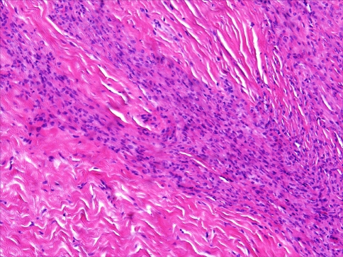
The perineurioma is an infrequently encountered benign peripheral nerve sheath tumor composed of a clonal proliferation of perineurial cells. Rare cases of perineurioma have been reported in the oral cavity. An extraneural sclerosing perineurioma arising in the buccal mucosa of a 17-year-old male is presented. Histopathologically, the tumor is composed of a well circumscribed nodular proliferation of spindle cells arranged in a storiform growth pattern, in some areas subtly arranged around vascular channels. The tumor cells reveal positive immunostaining for epithelial membrane antigen (EMA), collagen type IV and vimentin, and negative immunostaining for S-100 protein, consistent with a perineurial origin. To the best of our knowledge, this case represents the first report of an extraneural sclerosing perineurioma involving the oral cavity.


Figures

Fig. 1
Fig. 1
Low-power view showing a well-circumscribed densely collagenized proliferation of spindle cells exhibiting a vague storiform arrangement. (Hematoxylin and eosin [H&E], original magnification, 20×)
Fig. 2
Fig. 2
Hypercellular areas composed of spindle-shaped mesenchymal cells juxtaposed with hypocellular areas (H&E, original magnification, 100×)
Fig. 3
Fig. 3
Photomicrographs showing positive immunohistochemical staining in the tumor cell population for: a epithelial membrane antigen (EMA); b vimentin; and c collagen type IV. (Original magnification of each photomicrograph, 200×)

References

    1. Lazarus SS, Trombetta LD. Ultrastructural identification of a benign perineurial cell tumor. Cancer. 1978;41(5):1823–1829. doi: 10.1002/1097-0142(197805)41:5<1823::AID-CNCR2820410525>3.0.CO;2-J. - DOI - PubMed
    1. Fetsch JF, Miettinen M. Sclerosing perineurioma: a clinicopathologic study of 19 cases of a distinctive soft tissue lesion with a predilection for the fingers and palms of young adults. Am J Surg Pathol. 1997;21(12):1433–1442. doi: 10.1097/00000478-199712000-00005. - DOI - PubMed
    1. Barrett AW, Hopper C, Landon G. Intra-osseous soft tissue perineurioma of the inferior alveolar nerve. Oral Oncol. 2002;38(8):793–796. doi: 10.1016/S1368-8375(02)00005-2. - DOI - PubMed
    1. Boyanton BL, Jr, Jones JK, Shenaq SM, Hicks MJ, Bhattacharjee MB. Intraneural perineurioma: a systematic review with illustrative cases. Arch Pathol Lab Med. 2007;131(9):1382–1392. - PubMed
    1. da Cruz Perez DE, Amanajas de Aguiar FC, Jr, Leon JE, Graner E, Paes de Almeida O, Vargas PA. Intraneural perineurioma of the tongue: a case report. J Oral Maxillofac Surg. 2006;64(7):1140–1142. doi: 10.1016/j.joms.2006.03.032. - DOI - PubMed

Publication types