Review
. 2010 Oct;51(5):404-17.
doi: 10.3109/03008200903329789.

Dentin sialophosphoprotein in biomineralization

Affiliations
Review

Dentin sialophosphoprotein in biomineralization

Monica Prasad et al. Connect Tissue Res. 2010 Oct.

Abstract

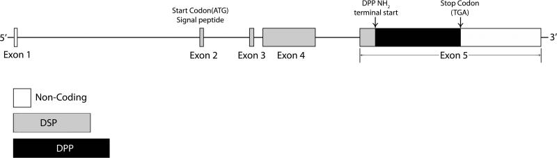
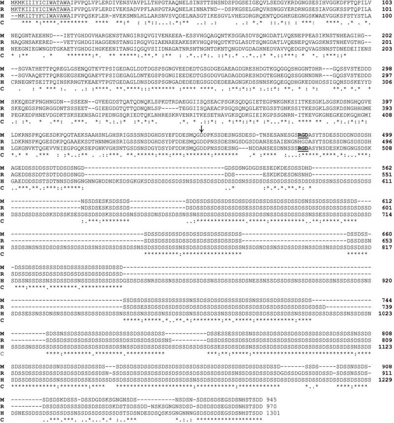
Two of the proteins found in significant quantity in the extracellular matrix (ECM) of dentin are dentin phosphoprotein (DPP) and dentin sialoprotein (DSP). DPP, the most abundant of the noncollagenous proteins (NCPs) in dentin is an unusually polyanionic protein, containing a large number of aspartic acids (Asp) and phosphoserines (Pse) in the repeating sequences of (Asp-Pse)(n). and (Asp-Pse-Pse)(n). The many negatively charged regions of DPP are thought to promote mineralization by binding calcium and presenting it to collagen fibers at the mineralization front during the formation of dentin. This purported role of DPP is supported by a sizeable pool of in vitro mineralization data showing that DPP is an important initiator and modulator for the formation and growth of hydroxyapatite (HA) crystals. Quite differently, DSP is a glycoprotein, with little or no phosphate. DPP and DSP are the cleavage products of dentin sialophosphoprotein (DSPP). Human and mouse genetic studies have demonstrated that mutations in, or knockout of, the Dspp gene result in mineralization defects in dentin and/or bone. The discoveries in the past 40 years with regard to DPP, DSP, and DSPP have greatly enhanced our understanding of biomineralization and set a new stage for future studies. In this review, we summarize the important and new developments made in the past four decades regarding the structure and regulation of the Dspp gene, the biochemical characteristics of DSPP, DPP, and DSP as well as the cell/tissue localizations and functions of these molecules.


Figures

Fig. 1
Fig. 1. Gene Structure of mouse DSPP
[20]. In the mouse DSPP gene, exon 1 contains a non-coding 5’ sequence; exon 2 contains the transcriptional start site, signal peptide, and first two amino acids of the NH2 -terminus of DSPP; exons 3 and 4 contain coding information for 29 and 314 amino acids, respectively; the remainder of the coding information and the untranslated 3’ region are contained in exon 5. The DSP sequence located at the NH2 terminus is found in exons 1–4 and the 5’ region of exon 5. The DPP sequence is located in the remainder of exon 5.
Fig. 2
Fig. 2. Comparison of amino acid sequences among mouse, rat and human DSPP
The amino acid sequences from mice [1], rats [25] and humans [26] have been aligned with the aid of the computer program Clustal W2 [88]. M, mouse; R, rat; H, human; C, consensus. The signal peptide region is underlined and the start of DPP (NH2-terminus) is denoted by a vertical arrow. The RGD motifs are underlined and bolded. “*” indicates that the residues in that column are identical in all sequences in the alignment. “:” indicates conserved substitutions. “.” indicates semi-conserved substitutions. “-” indicates that a deletion is observed compared with the other species. Blank spaces denote the absence of any substitution, indicating that these regions are not conserved.
Fig. 3
Fig. 3. Q-Sepharose separation and Stains-All staining for NCPs extracted from rat dentin by 4 M Gdm-HCl containing 0.5 M EDTA
Fig. 3A: NCPs were extracted from incisors of 12-week-old rats by 4 M Gdm-HCl containing 0.5 M EDTA and protease inhibitors (0.78 mg ml–1 of benzamidine-HCl, 0.18 mg ml–1 of sodium iodoacetate, 1.8 μg ml–1 of soybean trypsin inhibitor, 0.17 mg ml–1 of phenylmethylsufonyl fluoride, and 5 μg ml–1 of pepstatin). The extracts were separated by Q-Sepharose (Amersham Biosciences, Uppsala, Sweden) chromatography with a gradient ranging from 0.1–0.8 M NaCl in 6 M urea solution. This ion-exchange chromatography separated the NCPs into 95 fractions, each containing 1 ml of 6 M urea. Fig. 3B: Stains-All staining of fractions 22-92 from the Q-Sepharose chromatography. Numbers on the top of the figure represent fraction numbers. DSP is mainly present in fractions 24-36, appearing as a ~100 kDa blue band. DPP is primarily eluted in fractions 46-54, occurring as a very broad band, migrating around 100 kDa. The proteoglycan form of DSP is eluted in fractions 66-92, occurring as smears that are stained purple-blue.
Fig. 4
Fig. 4. Immunohistochemical staining of DSP in the first mandibular molar, alveolar bone and tibia of 5-week-old rats
Immunohistochemical staining was carried out using an ABC kit (Vector Laboratories Inc, Burlingame, CA, USA) following the manufacturer's instructions. The monoclonal anti-DSP antibody clone 2C12.3 [4] was used at a dilution of 1:1000 (approximately 1.9 μg ml-1 of IgG), and the same concentration of normal mouse IgG was used as a negative control. The sections were counterstained with methyl green (Fisher Scientific, Fair Lawn, NJ, USA) solution. Bar in Fig.4A: 100 μm; bars in Figs. 4B, 4C and 4D: 20 μm. Fig. 4A: Light microscopic images of the anti-DSP immunohistochemistry showing the buccolingual section of first mandibular molar, along with its surrounding periodontal tissues from a postnatal 5-week-old rat. D denotes dentin; DP indicates dental pulp. Arrow indicates cellular cementum (positive to the anti-DSP antibody). Arrowheads indicate alveolar bone. Note that intense reactions are detected in the alveolar bone at the bottom of the socket. Fig. 4B: High magnification of the boxed area in Fig. 4A. Note the presence of DSP in the matrix as well as in the osteocytes. Fig. 4C: Very weak positive reaction (arrows) to the anti-DSP antibody is observed in the cortical bone of the rat tibia. Please note that the immunostaining intensity of DSP is much weaker in the tibia than in the alveolar bone, suggesting that the expression level of DSP (DSPP) in the long bone is much lower than in the alveolar bone. Fig. 4D: Negative control. This section was serial to that used in Fig. 4C. Normal mouse IgG (1.9 μg ml-1) was used to replace the anti-DSP monoclonal antibody in the immunohistochemical staining protocol.

Similar articles

Cited by

References

    1. MacDougall M, Simmons D, Luan X, Nydegger J, Feng J, Gu TT. Dentin phosphoprotein and dentin sialoprotein are cleavage products expressed from a single transcript coded by a gene on human chromosome 4. Dentin phosphoprotein DNA sequence determination. J Biol Chem. 1997;272:835–42. - PubMed
    1. Fisher LW, Torchia DA, Fohr B, Young MF, Fedarko NS. Flexible structures of SIBLING proteins, bone sialoprotein, and osteopontin. Biochem Biophys Res Commun. 2001;280:460–5. - PubMed
    1. Qin C, Brunn JC, Cadena E, Ridall A, Tsujigiwa H, Nagatsuka H, Nagai N, Butler WT. The expression of dentin sialophosphoprotein gene in bone. J Dent Res. 2002;81:392–4. - PubMed
    1. Baba O, Qin C, Brunn JC, Jones JE, Wygant JN, McIntyre BW, Butler WT. Detection of dentin sialoprotein in rat periodontium. Eur J Oral Sci. 2004;112:163–70. - PubMed
    1. Alvares K, Kanwar YS, Veis A. Expression and potential role of dentin phosphophoryn (DPP) in mouse embryonic tissues involved in epithelial-mesenchymal interactions and branching morphogenesis. Dev Dyn. 2006;235:2980–90. - PubMed

Publication types

MeSH terms

LinkOut - more resources