

Sonic Hedgehog influences the balance of osteogenesis and adipogenesis in mouse adipose-derived stromal cells
- PMID: 20367246
- PMCID: PMC2947454
- DOI: 10.1089/ten.TEA.2010.0048
Sonic Hedgehog influences the balance of osteogenesis and adipogenesis in mouse adipose-derived stromal cells
Abstract
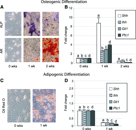
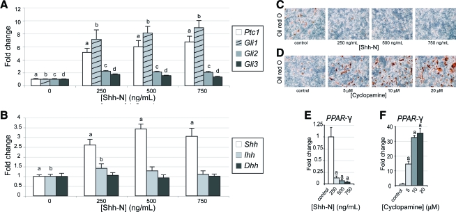
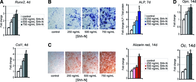
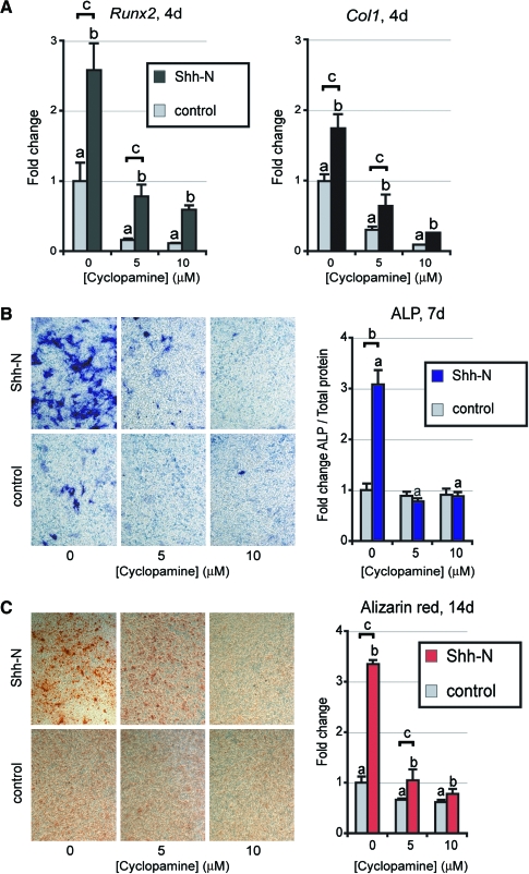
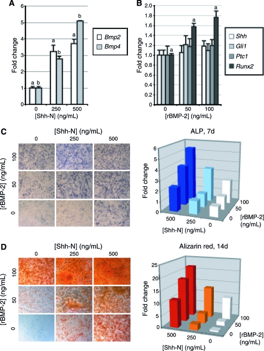
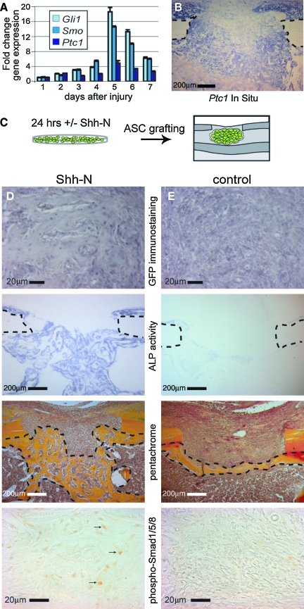
Adipose-derived stromal cells (ASCs) present a great potential for tissue engineering, as they are capable of differentiating into osteogenic and adipogenic cell types, among others. In this study, we examined the role of Hedgehog signaling in the balance of osteogenic and adipogenic differentiation in mouse ASCs. Results showed that Hedgehog signaling increased during early osteogenic differentiation (Shh, Ptc1, and Gli1), but decreased during adipogenic differentiation. N-terminal Sonic Hedgehog (Shh-N) significantly increased in vitro osteogenic differentiation in mouse ASCs, by all markers examined (*p < 0.01). Concomitantly, Shh-N abrogated adipogenic differentiation, by all markers examined (*p < 0.01). Conversely, blockade of endogenous Hedgehog signaling, with the Hedgehog antagonist cyclopamine, enhanced adipogenesis at the expense of osteogenesis. We next translated these results to a mouse model of appendicular skeletal regeneration. Using quantitative real-time polymerase chain reaction and in situ hybridization, we found that skeletal injury (a monocortical 1 mm defect in the tibia) results in a localized increase in Hedgehog signaling. Moreover, grafting of ASCs treated with Shh-N resulted in significantly increased bone regeneration within the defect site. In conclusion, Hedgehog signaling enhances the osteogenic differentiation of mouse ASCs, at the expense of adipogenesis. These data suggest that Hedgehog signaling directs the lineage differentiation of mesodermal stem cells and represents a promising strategy for skeletal tissue regeneration.
Figures
Similar articles
-
Human adipose-derived stromal cells stimulate autogenous skeletal repair via paracrine Hedgehog signaling with calvarial osteoblasts.Stem Cells Dev. 2011 Feb;20(2):243-57. doi: 10.1089/scd.2010.0250. Epub 2010 Oct 12. Stem Cells Dev. 2011. PMID: 20698749 Free PMC article.
-
Additive effects of sonic hedgehog and Nell-1 signaling in osteogenic versus adipogenic differentiation of human adipose-derived stromal cells.Stem Cells Dev. 2012 Aug 10;21(12):2170-8. doi: 10.1089/scd.2011.0461. Epub 2012 Feb 22. Stem Cells Dev. 2012. PMID: 22264144 Free PMC article.
-
Effect of 20(S)-Hydroxycholesterol on Multilineage Differentiation of Mesenchymal Stem Cells Isolated from Compact Bones in Chicken.Genes (Basel). 2020 Nov 17;11(11):1360. doi: 10.3390/genes11111360. Genes (Basel). 2020. PMID: 33213081 Free PMC article.
-
The role of microRNAs in cell fate determination of mesenchymal stem cells: balancing adipogenesis and osteogenesis.BMB Rep. 2015 Jun;48(6):319-23. doi: 10.5483/bmbrep.2015.48.6.206. BMB Rep. 2015. PMID: 25341923 Free PMC article. Review.
-
PPARγ and Wnt Signaling in Adipogenic and Osteogenic Differentiation of Mesenchymal Stem Cells.Curr Stem Cell Res Ther. 2016;11(3):216-25. doi: 10.2174/1574888x10666150519093429. Curr Stem Cell Res Ther. 2016. PMID: 25986621 Review.
Cited by
-
Primary cilia-mediated mechanotransduction in human mesenchymal stem cells.Stem Cells. 2012 Nov;30(11):2561-70. doi: 10.1002/stem.1235. Stem Cells. 2012. PMID: 22969057 Free PMC article.
-
Progressive osseous heteroplasia: diagnosis, treatment, and prognosis.Appl Clin Genet. 2015 Jan 30;8:37-48. doi: 10.2147/TACG.S51064. eCollection 2015. Appl Clin Genet. 2015. PMID: 25674011 Free PMC article. Review.
-
Human adipose-derived stromal cells stimulate autogenous skeletal repair via paracrine Hedgehog signaling with calvarial osteoblasts.Stem Cells Dev. 2011 Feb;20(2):243-57. doi: 10.1089/scd.2010.0250. Epub 2010 Oct 12. Stem Cells Dev. 2011. PMID: 20698749 Free PMC article.
-
Molecular analysis and differentiation capacity of adipose-derived stem cells from lymphedema tissue.Plast Reconstr Surg. 2013 Sep;132(3):580-589. doi: 10.1097/PRS.0b013e31829ace13. Plast Reconstr Surg. 2013. PMID: 23985633 Free PMC article.
-
BBS4 regulates the expression and secretion of FSTL1, a protein that participates in ciliogenesis and the differentiation of 3T3-L1.Sci Rep. 2017 Aug 29;7(1):9765. doi: 10.1038/s41598-017-10330-0. Sci Rep. 2017. PMID: 28852127 Free PMC article.
References
-
- Xu Y. Malladi P. Wagner D.R. Longaker M.T. Adipose-derived mesenchymal cells as a potential cell source for skeletal regeneration. Curr Opin Mol Ther. 2005;7:300. - PubMed
-
- Nuttall M.E. Gimble J.M. Controlling the balance between osteoblastogenesis and adipogenesis and the consequent therapeutic implications. Curr Opin Pharmacol. 2004;4:290. - PubMed
Publication types
MeSH terms
Substances
Grants and funding
LinkOut - more resources
Full Text Sources
