Review
. 2010:33:349-78.
doi: 10.1146/annurev-neuro-060909-153204.

Mechanisms of synapse and dendrite maintenance and their disruption in psychiatric and neurodegenerative disorders

Affiliations
Review

Mechanisms of synapse and dendrite maintenance and their disruption in psychiatric and neurodegenerative disorders

Yu-Chih Lin et al. Annu Rev Neurosci. 2010.

Abstract

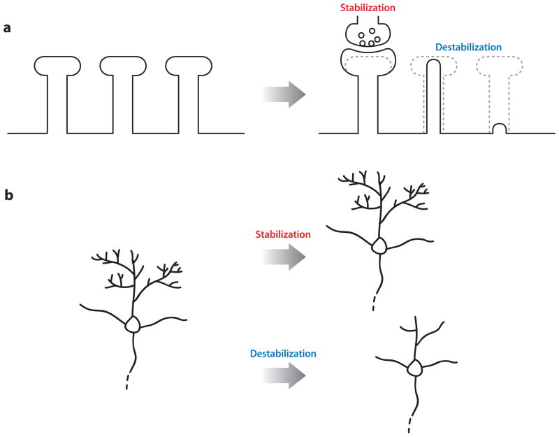
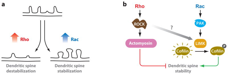
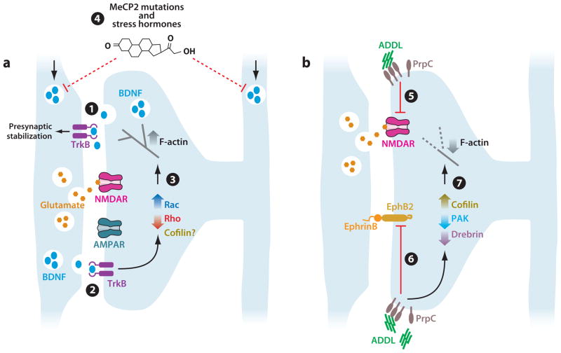
Emerging evidence indicates that once established, synapses and dendrites can be maintained for long periods, if not for the organism's entire lifetime. In contrast to the wealth of knowledge regarding axon, dendrite, and synapse development, we understand comparatively little about the cellular and molecular mechanisms that enable long-term synapse and dendrite maintenance. Here, we review how the actin cytoskeleton and its regulators, adhesion receptors, and scaffolding proteins mediate synapse and dendrite maintenance. We examine how these mechanisms are reinforced by trophic signals passed between the pre- and postsynaptic compartments. We also discuss how synapse and dendrite maintenance mechanisms are compromised in psychiatric and neurodegenerative disorders.


Figures

Figure 1
Figure 1
Stabilization of dendritic spines. (a) Synaptic activity, including adhesive contact between pre- and postsynaptic compartments and trophic signaling between the two compartments, enhances dendritic spine stability. (b) Synaptic support maintains the integrity of dendritic architecture over time. The loss of this support results in destabilization of dendritic spines followed by dendritic simplification.
Figure 2
Figure 2
Molecular mechanisms that regulate dendritic spine stability. The actin cytoskeleton and its regulators, adhesion receptors, and scaffolding proteins provide physical support for long-term synaptic maintenance. Most signaling pathways regulate spine stability by either 1. directly or 2. indirectly (via RhoGTPase signaling pathways) regulating actin dynamics or its interactions with adhesion and scaffolding molecules. 3. Adhesion molecules mediate signaling from the presynaptic or extracellular compartments into dendritic spines to evoke changes in Rho GTPase and other signaling cascades that control F-actin structure and stability. 4. Scaffolding proteins both interact directly with F-actin to support cytoskeletal structure and organize signaling molecules that impinge on these cytoskeletal control mechanisms.
Figure 3
Figure 3
Rho family GTPases have opposite effects on dendritic spine stability. (a) Rho and Rac have opposite effects on dendritic spine maintenance. Rho activation causes dendritic spine destabilization characterized by spine retraction and collapse, resulting in dendritic segments that lack spines. Rac activation promotes dendritic spine enlargement and stabilization. (b) Activation of RhoA stimulates Rho-associated protein kinase (ROCK) and actomyosin contractility, which causes spine collapse and synapse loss. Conversely, Rac activates Pak1 and LIMK to phosphorylate and inhibit cofilin, thereby stabilizing F-actin and promoting dendritic spine stability. Rho signaling through ROCK can also activate LIMK, but whether this contributes to spine stability is unclear.
Figure 4
Figure 4
Targeting of synaptic stabilization mechanisms in disease. (a) Model for synapse stabilization by BDNF signaling through TrkB. 1. Synaptic activity stimulates BDNF release from the postsynaptic compartment, which binds to presynaptic TrkB receptors. This leads to stabilization of the presynaptic compartment. 2. Activity-based BDNF release from the presynaptic compartment activates postsynaptic TrkB. 3. Postsynaptic TrkB may activate Rac, inhibit Rho, and regulate cofilin to stimulate increased F-actin assembly to stabilize synapses. 4. Mutations in the MeCP2 gene, as in Rett syndrome, and elevated stress hormones likely destabilize synapses by reducing BDNF levels. (b) Model for synapse destabilization by Aβ-derived diffusible ligands. 5, 6. Binding of ADDL (Aβ-derived diffusible ligands) to its receptor PrpC on the dendritic spine leads to reduced amounts of both NMDA receptor (5) and EphB2 (6), both of which help stabilize synapses. 7. ADDL stimulation increases cofilin activity, decreases PAK activity, and reduces drebrin levels leading to reduced F-actin levels and spine shrinkage.

Similar articles

Cited by

References

    1. Abe K, Chisaka O, Van Roy F, Takeichi M. Stability of dendritic spines and synaptic contacts is controlled by alpha N-catenin. Nat Neurosci. 2004;7:357–63. - PubMed
    1. Ackermann M, Matus A. Activity-induced targeting of profilin and stabilization of dendritic spine morphology. Nat Neurosci. 2003;6:1194–200. - PubMed
    1. Allison DW, Gelfand VI, Spector I, Craig AM. Role of actin in anchoring postsynaptic receptors in cultured hippocampal neurons: differential attachment of NMDA versus AMPA receptors. J Neurosci. 1998;18:2423–36. - PMC - PubMed
    1. Alvarez VA, Ridenour DA, Sabatini BL. Distinct structural and ionotropic roles of NMDA receptors in controlling spine and synapse stability. J Neurosci. 2007;27:7365–76. - PMC - PubMed
    1. Anderton BH, Callahan L, Coleman P, Davies P, Flood D, et al. Dendritic changes in Alzheimer’s disease and factors that may underlie these changes. Prog Neurobiol. 1998;55:595–609. - PubMed

Publication types

MeSH terms