Review
. 2010 Oct;13(8):1231-46.
doi: 10.1089/ars.2010.3187.

Mechanisms of oxidative protein folding in the bacterial cell envelope

Affiliations
Review

Mechanisms of oxidative protein folding in the bacterial cell envelope

Hiroshi Kadokura et al. Antioxid Redox Signal. 2010 Oct.

Abstract

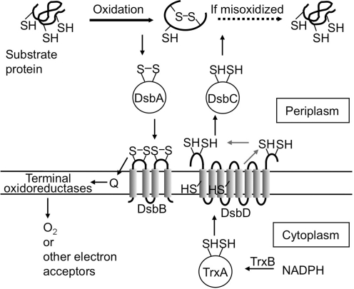
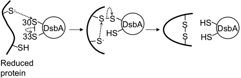
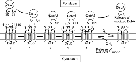
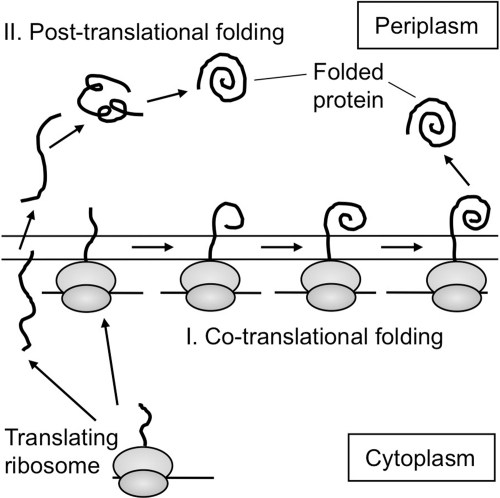
Disulfide-bond formation is important for the correct folding of a great number of proteins that are exported to the cell envelope of bacteria. Bacterial cells have evolved elaborate systems to promote the joining of two cysteines to form a disulfide bond and to repair misoxidized proteins. In the past two decades, significant advances have occurred in our understanding of the enzyme systems (DsbA, DsbB, DsbC, DsbG, and DsbD) used by the gram-negative bacterium Escherichia coli to ensure that correct pairs of cysteines are joined during the process of protein folding. However, a number of fundamental questions about these processes remain, especially about how they occur inside the cell. In addition, recent recognition of the increasing diversity among bacteria in the disulfide bond-forming capacity and in the systems for introducing disulfide bonds into proteins is raising new questions. We review here the marked progress in this field and discuss important questions that remain for future studies.


Figures

FIG. 1.
FIG. 1.
Models for disulfide-bond formation in the periplasm of E. coli. The solid black rightward arrow indicates the oxidative folding reaction catalyzed by DsbA. In the instance in which the first folding reaction resulted in a misoxidized protein, DsbC may repair it by acting as a reductase (dotted arrow) or an isomerase (not shown in this figure) of the incorrect disulfide bond. The thinner arrows indicate the flow of reducing equivalents. Q, quinones (ubiquinone or menaquinone). For simplicity, DsbC, a dimeric molecule, is depicted as a monomer.
FIG. 2.
FIG. 2.
The three-dimensional structure of DsbA. (A) The structure of the oxidized form of DsbA (PDB:1FVK) (74). DsbA has a thioredoxin-like fold with the insertion of an α-helical domain. The α-helices (blue), β-sheets (red), cis-Pro151 (magenta), and the sulfur atoms of the active-site cysteines (yellow) are indicated. Only the sulfur of Cys30 is surface-exposed. (B) The close-up view of the active site of the reduced form of DsbA (PDB: 1A2L) (33) in stick presentation. Possible hydrogen-bond interactions stabilizing the thiolate anion of Cys30 are indicated by dotted lines, with their distances in ångströms. A green ball represents the sulfur of Cys30. The distance between the sulfur of Cys30 and the main-chain oxygen of Val150 in the cis-Pro loop is also shown in ångströms with a solid line. Molecular graphics images were produced by using the UCSF Chimera (85). (For interpretation of the references to color in this figure legend, the reader is referred to the web version of this article at www.liebertonline.com/ars).
FIG. 3.
FIG. 3.
Disulfide-bond formation by DsbA. Substrate oxidation by DsbA likely proceeds through two steps. First, a deprotonated cysteine of a substrate attacks the sulfur atom of Cys30 of the oxidized DsbA, leading to the formation of a disulfide-linked complex between DsbA and the substrate. In the next step, one of the remaining cysteines of the substrate is deprotonated and attacks the sulfur atom of the substrate cysteine that is disulfide bonded with Cys30 of DsbA. This reaction results in the formation of a disulfide bond in the substrate and reduction of DsbA.
FIG. 4.
FIG. 4.
A model for early steps in the reoxidation of DsbA by DsbB. DsbA is highly oxidizing. Thus, it tends to be reduced. The mechanism by which DsbB oxidizes such a highly oxidizing protein is a fundamental question. In this model, the interloop disulfide bond formed between Cys130 and Cys41 (stage 4) prevents the back reaction (stage 2 to stage 0) that would tend to occur because of the highly oxidizing nature of DsbA. Thus, DsbB coordinates the action of its four cysteines to oxidize DsbA. In this model, the DsbB–DsbA complex in stage 4 corresponds to the charge-transfer complex that is depicted in Fig. 5. In the alternative model, electron transfer from DsbA to quinone proceeds through a sequential set of two thiol-disulfide exchange reactions, and then reduction of quinones through a charge-transfer complex (not shown). Q indicates the position of ubiquinone bound to DsbB.
FIG. 5.
FIG. 5.
A model for the oxidation of the N-terminal cysteine pair of DsbB by DsbB-bound ubiquinone. See text for the description of the model.
FIG. 6.
FIG. 6.
Disulfide-bond isomerization (A) and reduction (B) by DsbC. Two mechanisms have been proposed for the repair of a misoxidized cysteine pair by DsbC. After the reduction of the substrate protein in sequence (B), DsbA can reoxidize the substrate, potentially generating the correct disulfide bond (not shown).
FIG. 7.
FIG. 7.
The crystal structures of DsbC (PDB:1EEJ) (A) and DsbG (PDB:1V58) (B). DsbC and DsbG are V-shaped homodimers; each arm is a monomer containing an N-terminal dimerization domain and a C-terminal thioredoxin domain. The two domains are connected by a linker helix. The yellow spheres represent the sulfurs of active-site cysteines. The dimer form of DsbG was generated by applying crystallographic symmetry (85). (For interpretation of the references to color in this figure legend, the reader is referred to the web version of this article at www.liebertonline.com/ars).
FIG. 8.
FIG. 8.
A model for the disulfide-linked DsbA-substrate complex that forms during the cotranslational folding of PhoA. PhoA has two pairs of disulfide bonds. During cotranslational folding of PhoA, the PhoA Cys286 attacked DsbA to form a disulfide-linked complex (this model) between DsbA and the elongating chain of PhoA. This reaction took place when the PhoA polypeptide was elongated to ∼43 kDa in length. Further elongation of the polypeptide allowed Cys336 to attack the mixed-disulfide bond, completing the formation of the C-terminal disulfide bond of PhoA (not shown) (53).
FIG. 9.
FIG. 9.
Cotranslational and posttranslational oxidative folding of PhoA. Results (53) suggest that the mode of translocation (cotranslational versus posttranslational) can affect the folding process of a protein in the periplasm.

Similar articles

Cited by

References

    1. Alksne LE. Rasmussen BA. Dsb-insensitive expression of CcrA, a metallo-β-lactamase from Bacteroides fragilis, in Escherichia coli after amino acid substitution at two cysteine residues within CcrA. J Bacteriol. 1996;178:4306–4309. - PMC - PubMed
    1. Andersen CL. Matthey-Dupraz A. Missiakas D. Raina S. A new Escherichia coli gene, dsbG, encodes a periplasmic protein involved in disulphide bond formation, required for recycling DsbA/DsbB and DsbC redox proteins. Mol Microbiol. 1997;26:121–132. - PubMed
    1. Arredondo S. Segatori L. Gilbert HF. Georgiou G. De novo design and evolution of artificial disulfide isomerase enzymes analogous to the bacterial DsbC. J Biol Chem. 2008;283:31469–31476. - PMC - PubMed
    1. Arredondo SA. Chen TF. Riggs AF. Gilbert HF. Georgiou G. Role of dimerization in the catalytic properties of the Escherichia coli disulfide isomerase DsbC. J Biol Chem. 2009;284:23972–23979. - PMC - PubMed
    1. Åslund F. Berndt KD. Holmgren A. Redox potentials of glutaredoxins and other thiol-disulfide oxidoreductases of the thioredoxin superfamily determined by direct protein-protein redox equilibria. J Biol Chem. 1997;272:30780–30786. - PubMed

Publication types

MeSH terms

LinkOut - more resources