. 2010 Dec;19(12):1923-35.
doi: 10.1089/scd.2009.0326. Epub 2010 Oct 12.

Proliferation and pluripotency of human embryonic stem cells maintained on type I collagen

Affiliations

Proliferation and pluripotency of human embryonic stem cells maintained on type I collagen

Meredith B Jones et al. Stem Cells Dev. 2010 Dec.

Abstract

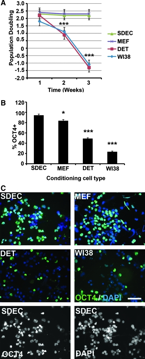
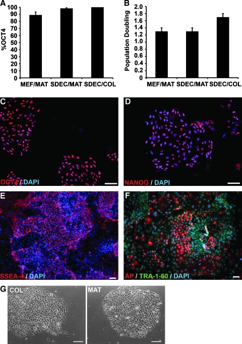
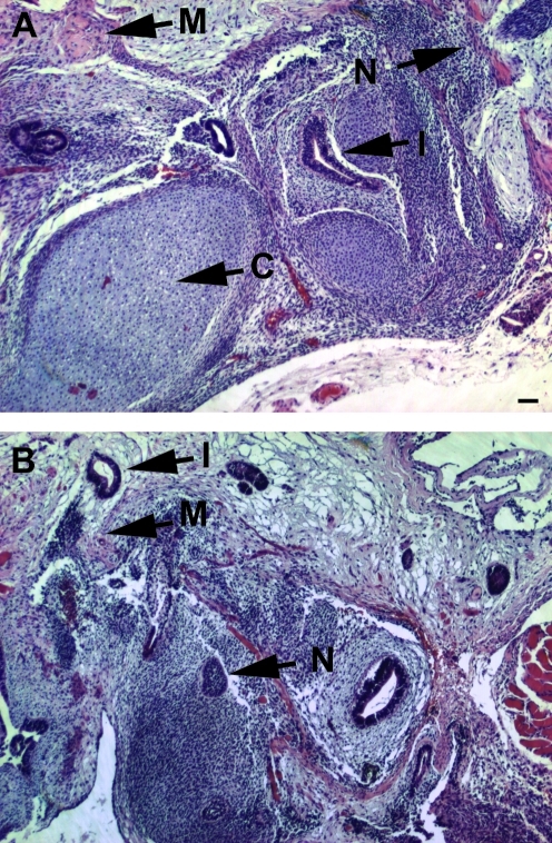
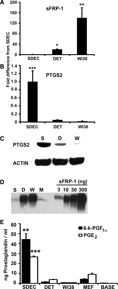
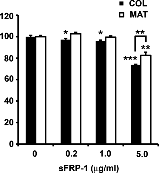
Human embryonic stem cells (hESC) require a balance of growth factors and signaling molecules to proliferate and retain pluripotency. Conditioned medium (CM) from a human embryonic germ-cell-derived cell culture, SDEC, was observed to support the growth of hESC on type I collagen (COL I) and on Matrigel (MAT) biomatricies. After 1 month, the population doubling of hESC grown in SDEC CM on COL I was equivalent to that of hESC grown in mouse embryonic fibroblast (MEF) CM on MAT. hESC grown in SDEC CM on COL I expressed OCT4, NANOG, SSEA-4, alkaline phosphatase (AP), and TRA-1-60; retained a normal karyotype; and were capable of forming teratomas. DNA microarray analysis was used to compare the transcriptional profiles of SDEC and the less supportive WI38 and Detroit 551 human cell lines. The mRNA level of secreted frizzled-related protein (sFRP-1), a known antagonist of the WNT/β-catenin signaling pathway, was significantly reduced in SDEC as compared with the other 2 cell lines, whereas the mRNA levels of prostaglandin-endoperoxide synthase 2 (PTGS2 or COX-2) and prostaglandin I₂ synthase (PGIS), two prostaglandin biosynthesis genes, were significantly increased in SDEC. The level of sFRP-1 protein was significantly reduced, and levels of 2 prostaglandins that are downstream products of PTGS2 and PGIS, prostaglandin E₂ and 6-keto-prostaglandin F(1α), were significantly elevated in SDEC CM compared with WI38, Detroit 551, and MEF CM. Further, addition of purified sFRP-1 to SDEC CM reduced the proliferation of hESC grown on COL I as well as MAT in a dose-dependent manner.


Figures

FIG. 1.
FIG. 1.
hESC proliferation and OCT4 expression for cells grown in SDEC-, MEF-, WI38-, and DET-conditioned medium on Matrigel. (A) Population doubling (mean ± SE, N = 3) of hESC over 3 weeks. (B) Percentage of OCT4-positive cells (mean ± SE, N = 3) after 3 weeks. (C) Immunofluorescent staining of hESC with OCT4 after 3 weeks. Nuclei stained with DAPI. Scale bar = 50 μm (magnification × 400). Significance: *P < 0.05 and ***P < 0.001. DAPI, 4′,6-diamidino-2-phenylindole; DET, Detroit 551; hESC, human embryonic stem cells; MEF, mouse embryonic fibroblast; SE, standard error.
FIG. 2.
FIG. 2.
hESC proliferation and expression of pluripotency markers for cells grown in SDEC or MEF CM on MAT or COL. (A) Percentage of OCT4-positive cells (mean ± SE, N = 4) after 1 month. (B) Population doubling (mean ± SE, N = 3) of hESC at the end of 1 month. (CF) Immunofluorescent staining of hESC grown in SDEC CM on COL after 1 month. Nuclei stained with DAPI. (C) OCT4, (D) NANOG, (E) SSEA-4, and (F) AP and TRA-1-60. (G) Phase microscopy of hESC colonies growing on COL and MAT. Scale bars = 50 μm (magnification × 400 for C, D, and G; magnification × 200 for E and F). AP, alkaline phosphatase; CM, conditioned medium; DAPI, 4′,6-diamidino-2-phenylindole; hESC, human embryonic stem cells; MAT, Matrigel; MEF, mouse embryonic fibroblast; SE, standard error.
FIG. 3.
FIG. 3.
Hematoxylin and eosin–stained sections of teratomas formed following transplantation of hESC into NOD/SCID mice. hESC lines (A) H1 and (B) H9. Tissue features marked: M, muscle; N, neuroectoderm; I, intestinal epithelia; C, cartilage. Scale bars = 100 μm (magnification × 100). hESC, human embryonic stem cells.
FIG. 4.
FIG. 4.
Expression of sFRP-1 and PTGS2 and prostaglandins by DET, WI38, and SDEC cell types. Quantitative RT-PCR was done to determine the fold difference in mRNA expression level (mean ± SE, N = 3) for (A) sFRP-1 and (B) PTGS2. mRNA levels were normalized using PPIA mRNA (housekeeping gene). Semiquantitative western blots were done on (C) intercellular PTGS2 (using β-actin to control for protein loading) and (D) sFRP-1 recovered from CM (180 mL from SDEC and DET, 150 mL from WI38). sFRP-1 recovered from MEF CM (180 mL) was also included in this western blot. The indicated amounts of recombinant human sFRP-1 were included for comparison. (E) Levels (mean ± SE, N = 3, ng/mL) of 6-k-PGF (black) and PGE2 (white) in CM from the 3 human cell types, MEF CM, and unconditioned human embryonic stem cells medium (BASE). Significance: **P < 0.01 and ***P < 0.001. CM, conditioned medium; DET, Detroit 551; MEF, mouse embryonic fibroblast; PGE2, prostaglandin E2, PTGS2, prostaglandin-endoperoxide synthase 2; RT-PCR, reverse transcription polymerase chain reaction; sFRP-1, secreted frizzled-related protein; 6-k-PGF, 6-keto-prostaglandin F; SE, standard error.
FIG. 5.
FIG. 5.
Cell proliferation assay of H9 human embryonic stem cells grown on COL or MAT matrices in SDEC-conditioned medium supplemented with various concentrations of sFRP-1. Proliferation of control (no added sFRP-1) was set to 100% and the percent of control proliferation (mean ± standard error, N = 4) for each sFRP-1 concentration is reported. Significance: *P < 0.05, **P < 0.01, and ***P < 0.001. MAT, Matrigel; sFRP-1, secreted frizzled-related protein.

Similar articles

Cited by

References

    1. Odorico JS. Kaufman DS. Thomson JA. Multilineage differentiation from human embryonic stem cell lines. Stem Cells. 2001;19:193–204. - PubMed
    1. Ho HY. Li M. Potential application of embryonic stem cells in Parkinson's disease: drug screening and cell therapy. Regen Med. 2006;1:175–182. - PubMed
    1. Porat S. Dor Y. New sources of pancreatic beta cells. Curr Diab Rep. 2007;7:304–308. - PubMed
    1. Shamblott MJ. Clark GO. Cell therapies for type 1 diabetes mellitus. Expert Opin Biol Ther. 2004;4:269–277. - PubMed
    1. Scharfmann R. Alternative sources of beta cells for cell therapy of diabetes. Eur J Clin Invest. 2003;33:595–600. - PubMed

Publication types

MeSH terms