

Decreased expression of PinX1 protein is correlated with tumor development and is a new independent poor prognostic factor in ovarian carcinoma
- PMID: 20367640
- PMCID: PMC11159430
- DOI: 10.1111/j.1349-7006.2010.01560.x
Decreased expression of PinX1 protein is correlated with tumor development and is a new independent poor prognostic factor in ovarian carcinoma
Abstract
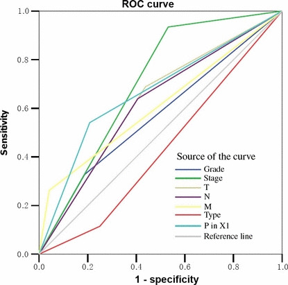
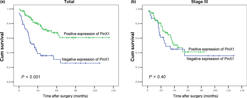
Human interacting protein X1 (PinX1) has been identified as a critical telomerase inhibitor and proposed to be a putative tumor suppressor gene. Loss of PinX1 has been found in a large variety of malignancies, but the expression status in epithelial ovarian tumors has not been investigated. In this study, immunohistochemistry for PinX1 protein was performed on a tissue microarray (TMA) of epithelial ovarian tumors (informatively containing 25 cystadenomas, 29 borderline tumors, and 157 invasive carcinomas) and 12 normal ovaries. Receiver-operator curve (ROC) analysis was used to determine cut-off scores for tumor positivity and to evaluate patients' survival status. The threshold for PinX1 positivity was determined to be above 60% (area under the curve = 0.856, P < 0.001) based on the area under the ROC. Positive expression of PinX1 was observed in 100% of normal ovarian tissues, in 84% of cystadenomas, in 75.9% borderline tumors, and 66.2% of ovarian carcinomas. Decreased expression of PinX1 was strongly related to patients with poor prognostic factors regarding presence of lymph node metastasis (P = 0.024), distant metastasis (P < 0.001), and late International Federation of Gynecology and Obstetrics (FIGO) stage (P < 0.001). In univariate survival analysis, a highly significant correlation between loss of PinX1 and shortened patient survival (mean, 48.2 months vs 99.2 months, P < 0.001) was displayed. Multivariate analysis demonstrated PinX1 expression (P = 0.027) was evaluated as an independent parameter. Our findings suggest that loss of PinX1 is an adverse independent molecular marker for epithelial ovarian carcinoma patients. PinX1 may be a novel target for telomerase-based anticancer therapy due to inhibiting telomerase activity.
Figures


Similar articles
-
PinX1 suppresses bladder urothelial carcinoma cell proliferation via the inhibition of telomerase activity and p16/cyclin D1 pathway.Mol Cancer. 2013 Nov 23;12(1):148. doi: 10.1186/1476-4598-12-148. Mol Cancer. 2013. PMID: 24268029 Free PMC article.
-
Significance of cell cycle regulatory proteins as malignant and prognostic biomarkers in ovarian epithelial tumors.Int J Gynecol Pathol. 2011 May;30(3):205-17. doi: 10.1097/PGP.0b013e3182063e71. Int J Gynecol Pathol. 2011. PMID: 21464733
-
PinX1 serves as a potential prognostic indicator for clear cell renal cell carcinoma and inhibits its invasion and metastasis by suppressing MMP-2 via NF-κB-dependent transcription.Oncotarget. 2015 Aug 28;6(25):21406-20. doi: 10.18632/oncotarget.4011. Oncotarget. 2015. PMID: 26033551 Free PMC article.
-
PinX1 plays multifaceted roles in human cancers: a review and perspectives.Mol Biol Rep. 2024 Nov 17;51(1):1163. doi: 10.1007/s11033-024-10082-x. Mol Biol Rep. 2024. PMID: 39550726 Free PMC article. Review.
-
The stromal origin of some epithelial ovarian neoplasms: "fere ex nihilo".Int J Gynecol Cancer. 2012 Jul;22(6):906-7. doi: 10.1097/IGC.0b013e31824ebba7. Int J Gynecol Cancer. 2012. PMID: 22740001 Review. No abstract available.
Cited by
-
The role of PinX1 in growth control of breast cancer cells and its potential molecular mechanism by mRNA and lncRNA expression profiles screening.Biomed Res Int. 2014;2014:978984. doi: 10.1155/2014/978984. Epub 2014 Feb 3. Biomed Res Int. 2014. PMID: 24672800 Free PMC article.
-
High expression of HEF1 predicts a poorer prognosis of hepatocellular carcinoma: A retrospective study.Mol Clin Oncol. 2016 Feb;4(2):159-165. doi: 10.3892/mco.2015.707. Epub 2015 Dec 11. Mol Clin Oncol. 2016. PMID: 26893853 Free PMC article.
-
Model based on five tumour immune microenvironment-related genes for predicting hepatocellular carcinoma immunotherapy outcomes.J Transl Med. 2021 Jan 6;19(1):26. doi: 10.1186/s12967-020-02691-4. J Transl Med. 2021. PMID: 33407546 Free PMC article.
-
High expression of p300 is linked to aggressive features and poor prognosis of nasopharyngeal carcinoma.J Transl Med. 2012 May 30;10:110. doi: 10.1186/1479-5876-10-110. J Transl Med. 2012. PMID: 22647238 Free PMC article.
-
High Expression of p300 in Human Breast Cancer Correlates with Tumor Recurrence and Predicts Adverse Prognosis.Chin J Cancer Res. 2011 Sep;23(3):201-7. doi: 10.1007/s11670-011-0201-5. Chin J Cancer Res. 2011. PMID: 23467396 Free PMC article.
References
-
- Lynch HT, Casey MJ, Lynch J, White TE, Godwin AK. Genetics and ovarian carcinoma. Semin Oncol 1998; 25: 265–80. - PubMed
-
- Kosary CL. FIGO stage, histology, histologic grade, age and race as prognostic factors in determining survival for cancers of the female gynecological system: an analysis of 1973–87 SEER cases of cancers of the endometrium, cervix, ovary, vulva, and vagina. Semin Surg Oncol 1994; 10: 31–46. - PubMed
-
- Engel J, Eckel R, Schubert‐Fritschle G et al. Moderate progress for ovarian cancer in the last 20 years: prolongation of survival, but no improvement in the cure rate. Eur J Cancer 2002; 38: 2435–45. - PubMed
-
- Baffa R, Santoro R, Bullrich F, Mandes B, Ishii H, Croce CM. Definition and refinement of chromosome 8p regions of loss of heterozygosity in gastric cancer. Clin Cancer Res 2000; 6: 1372–7. - PubMed
Publication types
MeSH terms
Substances
LinkOut - more resources
Full Text Sources
Medical
