Editorial
. 2010 Apr;14(4):729-40.
doi: 10.1111/j.1582-4934.2010.01059.x. Epub 2010 Mar 26.

TELOCYTES - a case of serendipity: the winding way from Interstitial Cells of Cajal (ICC), via Interstitial Cajal-Like Cells (ICLC) to TELOCYTES

Editorial

TELOCYTES - a case of serendipity: the winding way from Interstitial Cells of Cajal (ICC), via Interstitial Cajal-Like Cells (ICLC) to TELOCYTES

L M Popescu et al. J Cell Mol Med. 2010 Apr.

Abstract

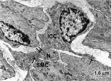
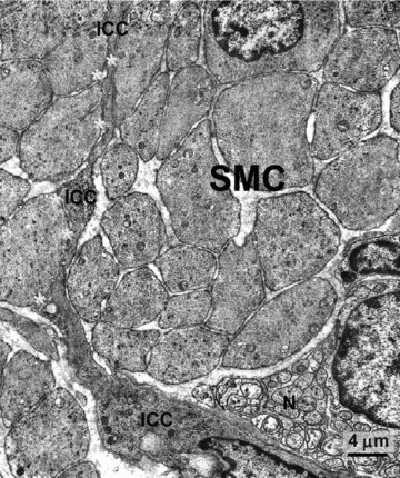
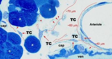
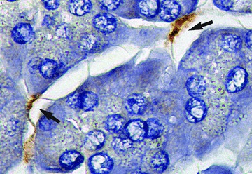
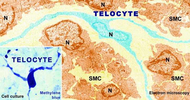
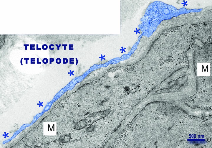
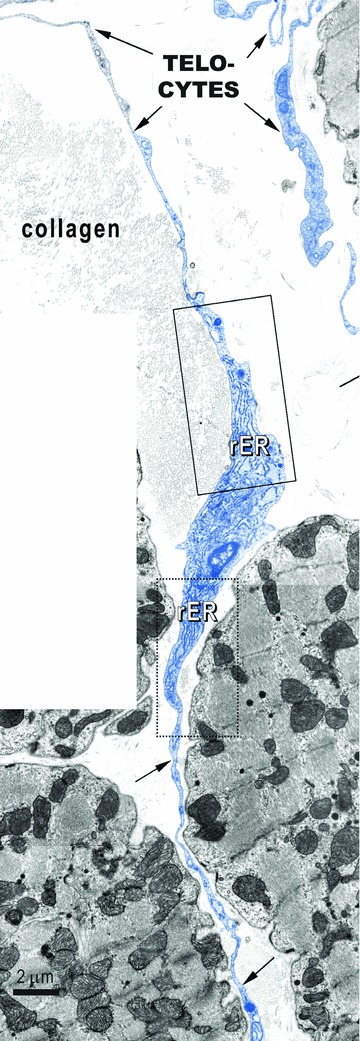
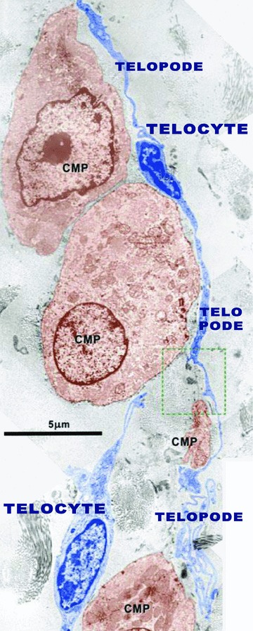
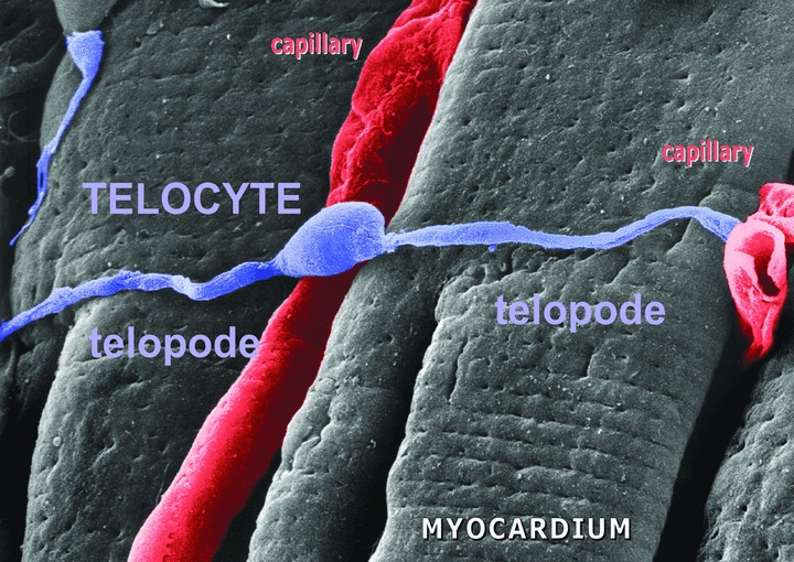
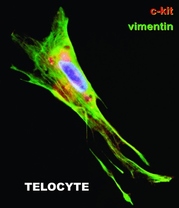
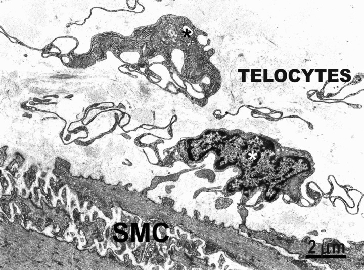
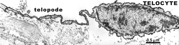
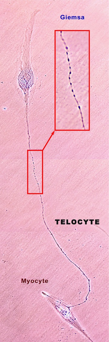
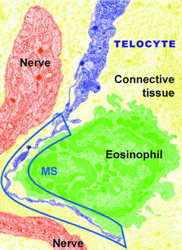
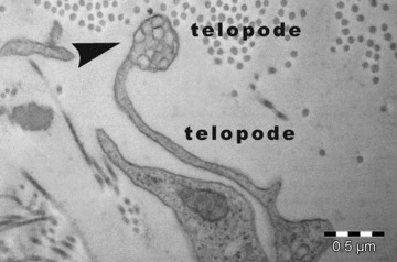
Ramon y Cajal discovered a particular cell type in the gut, which he named 'interstitial neurons' more that 100 years ago. In the early 1970s, electron microscopy/electron microscope (EM) studies showed that indeed a special interstitial cell type corresponding to the cells discovered by Cajal is localized in the gut muscle coat, but it became obvious that they were not neurons. Consequently, they were renamed 'interstitial cells of Cajal' (ICC) and considered to be pace-makers for gut motility. For the past 10 years many groups were interested in whether or not ICC are present outside the gastrointestinal tract, and indeed, peculiar interstitial cells were found in: upper and lower urinary tracts, blood vessels, pancreas, male and female reproductive tracts, mammary gland, placenta, and, recently, in the heart as well as in the gut. Such cells, now mostly known as interstitial Cajal-like cells (ICLC), were given different and confusing names. Moreover, ICLC are only apparently similar to canonical ICC. In fact, EM and cell cultures revealed very particular features of ICLC, which unequivocally distinguishes them from ICC and all other interstitial cells: the presence of 2-5 cell body prolongations that are very thin (less than 0.2 mum, under resolving power of light microscopy), extremely long (tens to hundreds of mum), with a moniliform aspect (many dilations along), as well as caveolae. Given the unique dimensions of these prolongations (very long and very thin) and to avoid further confusion with other interstitial cell types (e.g. fibroblast, fibrocyte, fibroblast-like cells, mesenchymal cells), we are proposing the term TELOCYTES for them, and TELOPODES for their prolongations, by using the Greek affix 'telos'.


Figures

Fig 1
Fig 1
Rabbit colon; circular muscle layer. One interstitial cell of Cajal (ICC) with an elongated body and three processes located in the connective interstitium. This cell has close cell-to-cell contact between both body and processes and two smooth muscle cells (SMC, arrows) and nerve endings (N, double arrows). This cell has the typical features of an intramuscular ICC: conspicuous Golgi apparatus and several rough endoplasmic reticulum cisternae; basal lamina is thin and discontinuous.
Fig 2
Fig 2
Human stomach; circular muscle layer. An interstitial cell of Cajal (ICC) located in the connective interstitium between the cross-sectioned bundles of smooth muscle cells (SMC), with which ICC establishes numerous cell-to-cell contacts (asterisks). Note the proximity of a large nerve bundle (N). The ICC body is spindle shaped and the cytoplasm is rich in filaments. The basal lamina is thick but discontinuous.
Fig 3
Fig 3
Rat exocrine pancreas. Non-conventional light microscopy; objective 100×. Tissue fixed with glutaraldehyde and post-fixed in OsO4. Thin section of Epon-embedded material (∼1 μm) was stained with toluidin blue; cap = capillary; ven = venule. At least four telocytes (TC) are present in the interstitium among acini (a). Note the cell bodies of TC and the emerging prolongations – telopodes (dashed lines). The length of the telopodes is very impressive: tens of micometres (!); they are very thin (less than 0.5 μm). Reproduced with permission from Popescu et al. [13].
Fig 4
Fig 4
Immunohistochemistry: Telocytes in human pancreas. Paraffin-embedded pancreas sections were incubated with polyclonal antibodies against CD 117. Nuclei were counterstained with Mayer haematoxylin. Telocytes (arrows) with fusiform body can be seen, having typical long, moniliform cytoplasmic processes that ‘touch’ the acini. Original magnification: 100×, oil immersion. Reproduced with permission from [12].
Fig 5
Fig 5
Human exocrine pancreas. Positive immunostaining of telocytes for CD34 (arrows), counterstained with Mayer’s haematoxylin, 40×. Reproduced with permission from [13].
Fig 6
Fig 6
Digitally coloured EM image of a telocyte in rat myometrium: telocyte (blue), SMCs (Sienna-brown); N = Nuclei. Note three long, moniliform processes that encircle bundles of cross-cut smooth muscle cells. Original magnification ×6800. Inset: Human pregnant myometrium. Primary confluent culture (day 8) showing a telocyte with at least 3 prolongations with several ‘beads’ along telopodes. Reproduced with permission from [14].
Fig 7
Fig 7
Electron microscopy of human non-pregnant uterus. Note the telocyte covering smooth muscle cells (M). The telopode is digitally coloured in blue, marked with asterisks. Image obtained in 2006. Courtesy of Prof. M. Taggart (Newcastle University, UK) and Dr. Carolyn J.P. Jones (Manchester University, UK).
Fig 8
Fig 8
EM image of telocytes (rat) in the right atrial interstitium; telocytes are indicated by arrows. Note the characteristic aspects of telopodes: very long and very thin cellular elongations, with uneven calibre (moniliform aspect). Rectangles show portions of telocyte body, containing (abundant) rough endoplasmic reticulum. Reproduced with permission from [30].
Fig 9
Fig 9
Electron micrograph from cardiac stem cells niche (sub-epicardium) illustrating the relationships of the telocyte (digitally blue coloured) with cardiomyocyte progenitors – CMP, (brown). The telocyte processes (telopodes) run parallel with the main axis of the CMP and seem to establish their direction of development. Reproduced with permission from [31].
Fig 10
Fig 10
Representative scanning electron micrograph. Monkey left ventricular myocardium. The image shows a typical telocyte located across the cardiomyocytes. Another (possible) telocyte appears located near the cardiomyocytes (upper left). The three-dimensional view reveals close interconnections of ICLCs with cardiomyocytes and capillaries (cap). Reproduced with permission from [30].
Fig 11
Fig 11
Human fallopian tube; subconfluent primary culture. Double immunofluorescent labelling of an ‘octopus’-like telocyte: vimentin (green) and CD117/ckit (red). Vimentin reactivity is mainly localized within the cell processes, and CD117/c-kit has a patchy pattern. The cell nucleus is shown in blue (Hoechst 33342); original magnification 60×. Reproduced with permission from [16].
Fig 12
Fig 12
Rabbit colon. Two telocytes (asterisks) close to the submucosal border of the circular muscle layer. These cells have a small oval body, mainly occupied by the nucleus, and extremely thin and long processes extend beyond the cellular body, curving repeatedly. No basal lamina is present around these cells. SMC – smooth muscle cells.
Fig 13
Fig 13
CD34-immunoelectro-labelling: small intestine. CD34-immunoelectro-labelling is present on the surface of a telocyte. The labelling appears as an electron-dense material distributed all along the plasma membrane, from which spherules protrude outside. Reproduced with kind permission from [52].
Fig 14
Fig 14
The growing scientific interest in ICLC (now TELOCYTES). Charts based on data released by Web of Science (Thomson Reuters ISI Web of Knowledge).
Fig 15
Fig 15
Human non-pregnant myometrium in cell culture; day 3; the first passage. Giemsa staining. A telocyte establishing contacts with a myocyte by a telopode of about 65 μm long. Photographic composition of 4 serial phase contrast images, original magnification 40×. In red rectangles, a higher magnification clearly shows the moniliform aspect; at least 40 specific dilations are visible in a ‘bead-like’ fashion. Reproduced with permission from [14].
Fig 16
Fig 16
Rat myometrium: TEM; original magnification 5700×; image digitally coloured. A multicontact synapse (MS) between an eosinophil (green) and a telocyte (violet) indicated by cassette. An unmyelinated nerve bundle (vermilion) appears in proximity of telocyte, but has no direct contact with it. Apparently, the telocyte ‘prefers’ the eosinophil. Synapse: type – multicontact synapse (MS); length – 8.64 μm; cleft width – 74 ± 26 nm; contact points – 13 (15 ± 8 nm wide). Reproduced with permission from [53].
Fig 17
Fig 17
Electron micrographs show details of telopodes from mouse epicardium. Shed vesicles (arrowhead) emerge from telopodes in the interstitial space.

Similar articles

Cited by

References

    1. Ramon y Cajal S. Histologie du Systeme Nerveux de L’Homme et des Vertebres. Vol. 2. Paris: A. Maloine; 1911.
    1. Faussone Pellegrini MS, Cortesini C, Romagnoli P. Ultrastructure of the tunica muscularis of the cardial portion of the human esophagus and stomach, with special reference to the so-called Cajal’s interstitial cells. Arch Ital Anat Embriol. 1977;82:157–77. - PubMed
    1. Thuneberg L. Interstitial cells of Cajal: intestinal pacemaker cells? Adv Anat Embryol Cell Biol. 1982;71:1–130. - PubMed
    1. Faussone-Pellegrini MS, Thuneberg L. Guide to the identification of interstitial cells of Cajal. Microsc Res Tech. 1999;47:248–66. - PubMed
    1. Faussone-Pellegrini MS. Interstitial cells of Cajal: once negligible players, now blazing protagonists. Ital J Anat Embryol. 2005;110:11–31. - PubMed

Publication types