. 2010 Apr 5:7:27.
doi: 10.1186/1742-4690-7-27.

Changes in the accessibility of the HIV-1 Integrase C-terminus in the presence of cellular proteins

Affiliations

Changes in the accessibility of the HIV-1 Integrase C-terminus in the presence of cellular proteins

Sofia Benkhelifa-Ziyyat et al. Retrovirology. .

Abstract

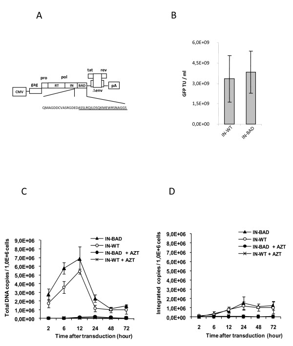
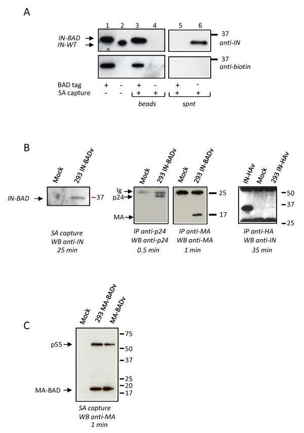
Background: Following entry, uncoating, and reverse transcription, a number of cellular proteins become associated with the human immunodeficiency virus type 1 (HIV-1) pre-integration complex (PIC). With the goal of obtaining reagents for the analysis of the HIV-1 PIC composition and localisation, we have constructed functional integrase (IN) and matrix (MA) proteins that can be biotinylated during virus production and captured using streptavidin-coated beads.

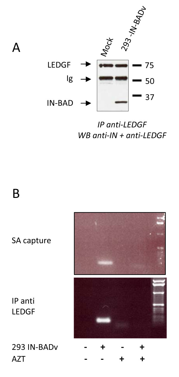
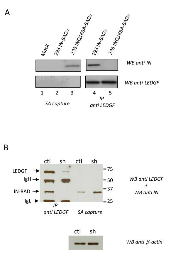
Results: Although the labelled C-terminus allows for the sensitive detection of virion-associated IN, it becomes inaccessible in the presence of cellular proteins. This masking is not dependent on the nature of the tag and does not occur with the tagged MA. It was not observed either with an IN mutant unable to interact with LEDGF/p75, or when LEDGF/p75 was depleted from cells.

Conclusion: Our observation suggests that a structural rearrangement or oligomerization of the IN protein occurs during the early steps of infection and that this process is related to the presence of LEDGF/p75.


Figures

Figure 1
Figure 1
Fusion of the Biotin Acceptor Domain (BAD) to the IN C-terminus does not affect particle production, cDNA synthesis, and integration. (A) Amino acid sequence at the C-terminus of IN-BAD, in the context of a p8.74 derived gagpol expression construct. (B) Comparison of vector titres obtained with IN-BAD and IN-WT. Data represent the mean ± SD of GFP titres measured on HCT116 cells from three independent productions. (C) Kinetics of HIV-1 vector DNA synthesis during vector transduction of HEK 293 cells (30 ng of p24gag/106 cells) with or without AZT, analysed by quantitative PCR. (D) Amounts of integrated provirus. Data in C and D represent the mean ± SD of three independent transductions.
Figure 2
Figure 2
IN-BAD is efficiently biotinylated in producer cells and incorporated into virions. IN-BAD (lanes 1, 3) or IN-WT (lanes 2, 4) vector particles (30 ng of p24gag) were either untreated (lanes 1, 2) or incubated with streptavidin paramagnetic beads and eluted (SA capture, lanes 3, 4). Samples were run on SDS-PAGE and Western blots (WB) were analysed with anti-IN (top) or anti-biotin (bottom) antibodies (1 minute exposure). Supernatants (spnt) from SA captures were also analysed (lane 5 and 6). (B) Left panel: streptavidin paramagnetic beads capture (SA capture) of the biotinylated IN (IN-BAD) from extracts of 293 cells mock-transduced (Mock) or transduced with the IN-BAD vector (293 IN-BADv), analysed by Western blotting with an anti-IN antibody. Middle panels: as controls, MA or p24 were immunoprecipitated (IP) respectively with an anti-MA and an anti p24 antibodies from the same cells extracts and analysed by WB respectively with the same antibodies. Right panel: HA tagged integrase (IN-HA) was immunoprecipitated with an anti-HA antibody from lysed IN-HA vector (IN-HAv) or from extracts of 293 cells mock-transduced (Mock) or transduced with IN-HAv (293 IN-HAv) and analysed by Western blotting with an anti-IN antibody. (C) Streptavidin paramagnetic beads capture of the biotinylated MA (MA-BAD) from extracts of 293 cells mock-transduced (Mock) or transduced with the MA-BAD vector (293 MA-BADv), or from lysed MA-BAD vector (MA-BADv) analysed by Western blotting with an anti-IN antibody.
Figure 3
Figure 3
(A) IN-BAD and LEDGF/p75 co-immunoprecipitation from extracts of 293 cells mock-transduced (Mock) or transduced with the IN-BADv (293 IN-BADv), analysed by Western blotting with anti-LEDGF/p75 and anti-IN antibodies (1 minute exposure). (B) PCR detection of viral DNA in streptavidin capture (top) and LEDGF/p75 immunoprecipitates (bottom). 293 cells were transduced with the IN-BAD vector (293 IN-BADv) or mock transduced (Mock) in the absence or presence of AZT. Streptavidine capture or LEDGF/p75 co-immunoprecipitation were performed on cell lysates, and vector DNA was detected using PCR with the MH531 and MH532 primers [17]. The absence of amplification in the presence of AZT indicates that only neo-synthesized DNA was detected.
Figure 4
Figure 4
Masking of the IN C-terminus in infected cells. (A) Streptavidin paramagnetic beads capture (SA capture) (1,2,3) or LEDGF/p75 co-immunoprecipitation (4,5) of the biotinylated IN from extracts of 293 cells mock-transduced (lane 1) or transduced with the IN-BAD vector (293 IN-BADv) (lane 2, 4) or INQ168A-BAD vector (293 INQ168A-BADv (lane 3, 5) analysed by Western blotting with the anti-IN (top) or anti LEDGF/p75 (bottom) antibodies (3 minutes exposure). (B) LEDGF/p75 co-immunoprecipitation or streptavidin capture of the biotinylated integrase from extracts of 293ctl (ctl) or 293sh (sh) mixed with the IN-BAD vector (IN-BADv). As a control, equal amount of 293ctl or 293sh lysates were tested for beta-actin content by WB (bottom panel).

Similar articles

Cited by

References

    1. Delelis O, Carayon K, Saib A, Deprez E, Mouscadet JF. Integrase and integration: biochemical activities of HIV-1 integrase. Retrovirology. 2008;5:114. doi: 10.1186/1742-4690-5-114. - DOI - PMC - PubMed
    1. Turlure F, Devroe E, Silver PA, Engelman A. Human cell proteins and human immunodeficiency virus DNA integration. Front Biosci. 2004;9:3187–3208. doi: 10.2741/1472. - DOI - PubMed
    1. Van Maele B, Busschots K, Vandekerckhove L, Christ F, Debyser Z. Cellular co-factors of HIV-1 integration. Trends Biochem Sci. 2006;31:98–105. doi: 10.1016/j.tibs.2005.12.002. - DOI - PubMed
    1. Llano M, Delgado S, Vanegas M, Poeschla EM. Lens Epithelium-derived Growth Factor/p75 Prevents Proteasomal Degradation of HIV-1 Integrase. J Biol Chem. 2004;279:55570–55577. doi: 10.1074/jbc.M408508200. - DOI - PubMed
    1. Mulder LCF, Muesing MA. Degradation of HIV-1 Integrase by the N-end Rule Pathway. J Biol Chem. 2000;275:29749–29753. doi: 10.1074/jbc.M004670200. - DOI - PubMed

Publication types

Substances