

Cholesterol granuloma presenting as a mass obstructing the external ear canal
- PMID: 20367883
- PMCID: PMC2853487
- DOI: 10.1186/1472-6815-10-4
Cholesterol granuloma presenting as a mass obstructing the external ear canal
Abstract
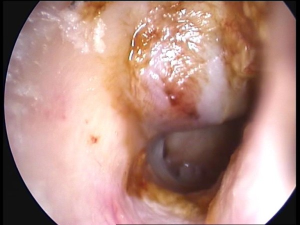
Background: Cholesterol granuloma (CG) may involve the middle ear, the mastoid bone and the petrous apex. However, CG presenting as a mass obstructing the external ear canal (EEC) is relatively rare and it can be a diagnostic challenge.
Case presentation: We report a case of a CG occupying the mastoid antrum and presenting as a mass into the EEC. Temporal bone computerized tomography showed a soft tissue mass which eroded the posterior-superior bony wall of the EEC. On magnetic resonance imaging, the mass revealed a high signal on both T1 and T2-weighted images. The CG was removed by a mastoidectomy procedure and the histopathologic report confirmed the diagnosis of CG. A type III tympanoplasty was performed.
Conclusions: The postoperative course was uneventful.
Figures


References
-
- Martin N, Sterkers O, Mompoint D, Julien N, Nahum H. Cholesterol Granulomas of the Middle Ear Cavities: MR Imaging. Radiology. 1989;172:521–525. - PubMed
-
- Rinaldo A, Ferlito A, Cureoglu S, Devaney K, Schachern P, Paparella M. Cholesterol granuloma of the temporal bone: a pathologic designation or a clinical diagnosis? Acta Oto-Laryngologica. 2005;125:186–190. - PubMed
-
- Campana D, Alves F, Sarmento P Neto, Palermo F, Salem L, Almeida P. Lesao litica do meato acustico externo granuloma de cholesterol. Rev Bras Otorrinolaringol. 1988;54:112–114.
-
- Koh S, Kim C, Lee S. Cholesterol granuloma of the external auditory canal. Korean J Otolaryngol. 2008;51:1143–1146.
LinkOut - more resources
Full Text Sources
Miscellaneous
