

Investigation of the direct effects of salmon calcitonin on human osteoarthritic chondrocytes
- PMID: 20367884
- PMCID: PMC2858096
- DOI: 10.1186/1471-2474-11-62
Investigation of the direct effects of salmon calcitonin on human osteoarthritic chondrocytes
Abstract
Background: Calcitonin has been demonstrated to have chondroprotective effects under pre-clinical settings. It is debated whether this effect is mediated through subchondral-bone, directly on cartilage or both in combination. We investigated possible direct effects of salmon calcitonin on proteoglycans and collagen-type-II synthesis in osteoarthritic (OA) cartilage.
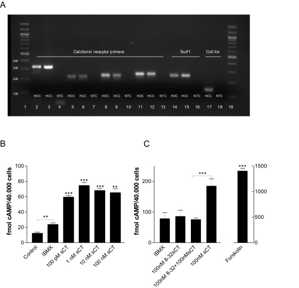
Methods: Human OA cartilage explants were cultured with salmon calcitonin [100 pM-100 nM]. Direct effects of calcitonin on articular cartilage were evaluated by 1) measurement of proteoglycan synthesis by incorporation of radioactive labeled 35SO4 [5 microCi] 2) quantification of collagen-type-II formation by pro-peptides of collagen type II (PIINP) ELISA, 3) QPCR expression of the calcitonin receptor in OA chondrocytes using four individual primer pairs, 4) activation of the cAMP signaling pathway by EIA and, 5) investigations of metabolic activity by AlamarBlue.
Results: QPCR analysis and subsequent sequencing confirmed expression of the calcitonin receptor in human chondrocytes. All doses of salmon calcitonin significantly elevated cAMP levels (P < 0.01 and P < 0.001). Calcitonin significantly and concentration-dependently [100 pM-100 nM] induced proteoglycan synthesis measured by radioactive 35SO4 incorporation, with a 96% maximal induction at 10 nM (P < 0.001) corresponding to an 80% induction of 100 ng/ml IGF, (P < 0.05). In alignment with calcitonin treatments [100 pM-100 nM] resulted in 35% (P < 0.01) increased PIINP levels.
Conclusion: Calcitonin treatment increased proteoglycan and collagen synthesis in human OA cartilage. In addition to its well-established effect on subchondral bone, calcitonin may prove beneficial to the management of joint diseases through direct effects on chondrocytes.
Figures


Similar articles
-
Evidence that human cartilage and chondrocytes do not express calcitonin receptor.Osteoarthritis Cartilage. 2008 Apr;16(4):450-7. doi: 10.1016/j.joca.2007.08.003. Epub 2007 Sep 24. Osteoarthritis Cartilage. 2008. PMID: 17890110
-
Calcitonin directly attenuates collagen type II degradation by inhibition of matrix metalloproteinase expression and activity in articular chondrocytes.Osteoarthritis Cartilage. 2006 Aug;14(8):759-68. doi: 10.1016/j.joca.2006.01.014. Epub 2006 Mar 20. Osteoarthritis Cartilage. 2006. PMID: 16549372
-
The inhibitory effect of salmon calcitonin on tri-iodothyronine induction of early hypertrophy in articular cartilage.PLoS One. 2012;7(6):e40081. doi: 10.1371/journal.pone.0040081. Epub 2012 Jun 29. PLoS One. 2012. PMID: 22768225 Free PMC article.
-
Calcitonin affects both bone and cartilage: a dual action treatment for osteoarthritis?Ann N Y Acad Sci. 2007 Nov;1117:181-95. doi: 10.1196/annals.1402.041. Ann N Y Acad Sci. 2007. PMID: 18056043 Review.
-
Calcitonin is involved in cartilage homeostasis: is calcitonin a treatment for OA?Osteoarthritis Cartilage. 2006 Jul;14(7):617-24. doi: 10.1016/j.joca.2006.03.014. Epub 2006 May 12. Osteoarthritis Cartilage. 2006. PMID: 16698291 Review.
Cited by
-
12-Deoxyphorbol-13-Hexadecanoate Abrogates OVX-Induced Bone Loss in Mice and Osteoclastogenesis via Inhibiting ROS Level and Regulating RANKL-Mediated NFATc1 Activation.Front Pharmacol. 2022 Jun 3;13:899776. doi: 10.3389/fphar.2022.899776. eCollection 2022. Front Pharmacol. 2022. PMID: 35721216 Free PMC article.
-
Impact of treatments for osteoporosis on cartilage biomarkers in humans.Osteoporos Int. 2012 Dec;23 Suppl 8:S877-80. doi: 10.1007/s00198-012-2165-9. Epub 2012 Nov 22. Osteoporos Int. 2012. PMID: 23179570 Review.
-
Altered AIB1 or AIB1Δ3 expression impacts ERα effects on mammary gland stromal and epithelial content.Mol Endocrinol. 2011 Apr;25(4):549-63. doi: 10.1210/me.2010-0114. Epub 2011 Feb 3. Mol Endocrinol. 2011. PMID: 21292825 Free PMC article.
-
The effects of calcitonin on post-orthodontic relapse in rats.Clin Exp Dent Res. 2021 Jun;7(3):293-301. doi: 10.1002/cre2.373. Epub 2020 Dec 9. Clin Exp Dent Res. 2021. PMID: 33300289 Free PMC article.
-
Oral calcitonin.Int J Womens Health. 2012;4:471-9. doi: 10.2147/IJWH.S24776. Epub 2012 Sep 6. Int J Womens Health. 2012. PMID: 23071417 Free PMC article.
References
Publication types
MeSH terms
Substances
LinkOut - more resources
Full Text Sources
Medical
