. 2009 Dec 2;1(9):9ra20.
doi: 10.1126/scitranslmed.3000284.

Identification of an autoantigen demonstrates a link between interstitial lung disease and a defect in central tolerance

Affiliations

Identification of an autoantigen demonstrates a link between interstitial lung disease and a defect in central tolerance

Anthony K Shum et al. Sci Transl Med. .

Abstract

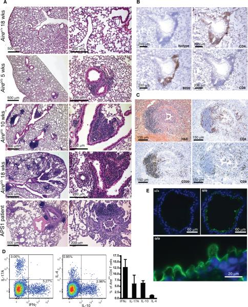
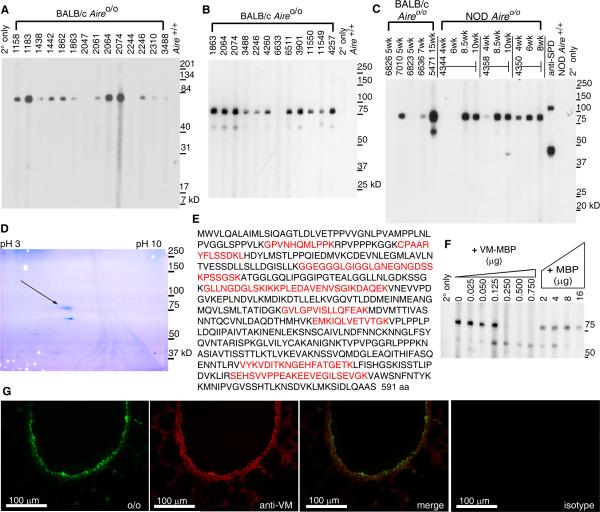
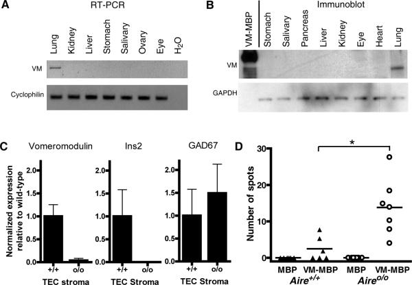
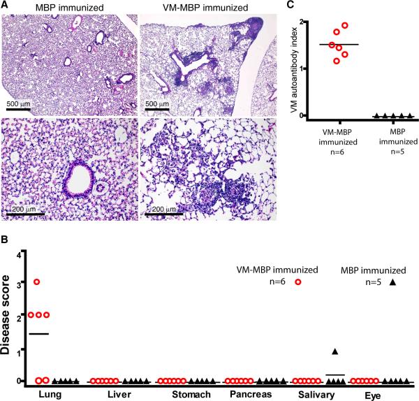
Interstitial lung disease (ILD) is a common manifestation of systemic autoimmunity characterized by progressive inflammation or scarring of the lungs. Patients who develop these complications can exhibit significantly impaired gas exchange that may result in hypoxemia, pulmonary hypertension, and even death. Unfortunately, little is understood about how these diseases arise, including the role of specific defects in immune tolerance. Another key question is whether autoimmune responses targeting the lung parenchyma are critical to ILD pathogenesis, including that of isolated idiopathic forms. We show that a specific defect in central tolerance brought about by mutations in the autoimmune regulator gene (Aire) leads to an autoreactive T cell response to a lung antigen named vomeromodulin and the development of ILD. We found that a human patient and mice with defects in Aire develop similar lung pathology, demonstrating that the AIRE-deficient model of autoimmunity is a suitable translational system in which to unravel fundamental mechanisms of ILD pathogenesis.


Figures

Fig. 1
Fig. 1. Pulmonary disease in Aireo/o mice and an APS1 patient
(A) Representative H&E of lungs from BALB/c Aireo/o and Aire+/+ mice. (Lower panels) H&E of lung biopsy from the APS1 patient. (B) Immunostaining of lungs from NOD Aireo/o mouse at 8 weeks for CD4, CD8, B cells and isotype control antibody. (C) Lung biopsy from an APS1 patient shows chronic bronchiolitis and prominent lymphoid aggregates. Immunostaining shows a nodular aggregate of B-cells (CD20+) consistent with early germinal center formation and scattered surrounding CD4+ and CD8+ T cells. (D) Representative plot of CD4+ lung lymphocytes from a BALB/c Aireo/o mouse at 14 weeks, showing IL-17A, IFNγ, IL-4 and IL-10 containing cells. (Right panel) Percentages of total CD4+ lung lymphocytes producing cytokines averaged from 5 BALB/c Aireo/o mice aged 12–16 weeks. Data are mean ± SEM. (E) Indirect immunofluorescence stain with serum from a NOD Aireo/o mouse with pulmonary disease on frozen lung section from immunodeficient SCID mouse. (Lower panel) Higher magnification image of lung section shown in upper panel, right. Green, serum staining; blue, staining with nuclear marker 4',6'-diamidino-2-phenylindole (DAPI).
Fig. 2
Fig. 2. Vomeromodulin, the predominant antigen targeted in lungs of Aireo/o mice
(A) Immunoblot of whole lung lysate probed with sera from BALB/c Aireo/o mice aged 8–20 weeks (individual animals numbered) revealed an 80 kD antigen target. (B) Immunoblot of BAL fluid probed with sera from BALB/c Aireo/o mice also revealed the 80 kD antigen. (C) Immunoblot of BAL fluid probed with NOD mice bled serially and BALB/c mice sacrificed at various ages. (D) Sera from Aireo/o mice were used to immunoprecipitate the antigen from BAL fluid, which was then run on a coomassiestained 2D gel. Three spots at 80 kD migrated near an isoelectric point ~5.5, (arrow). All spots were analyzed by mass spectrometry (E) The sequence of the 80 kD spot, indicating that it is vomeromodulin. Identified peptides (in red) mapped onto the VM amino acid sequence revealed coverage of nearly the entire protein. (F) To confirm autoantibody reactivity to VM, a competition blot showed 80 kD reactivity was abolished after addition of recombinant VM-MBP. The MBP tag alone failed to abolish reactivity. (G) Aireo/o serum and anti-VM sera co-localized in indirect immunofluorescence staining on lung tissue targeting the bronchiolar epithelial surface and less frequently, to cells within distal airways. SPD = surfactant protein D.
Fig. 3
Fig. 3. T cells with specificity for vomeromodulin in Aireo/o mice
(A) RT-PCR of vomeromodulin cDNA after 35 cycles in indicated tissues reveals a band at expected size of 1.8 kb in lung only. The DNA band was excised and sequenced, confirming that full length VM cDNA was amplified. (B) Immunoblot using BALB/c Aireo/o serum to probe tissue lysates of indicated organs and recombinant VM-MBP. Reactivity to the 80 kD band is only seen in lung lysate. Reactivity to VM-MBP occurs at expected weight of 100 kD. (C) Representative results from two independent experiments in which TEC stroma from Aireo/o and Aire+/+ thymi was assayed in quadruplicate for VM, insulin 2 (Ins2) and glutamic acid decarboxylase 67 (GAD67) by real-time PCR; data are normalized expression relative to wild-type ±SD. (D) ELISPOT analysis of IFNγ producing T cells in Aireo/o and Aire+/+ BALB/c mice aged 10–14 weeks (*P=.005). Y axis indicates number of spots per 10,000 T cells.
Fig. 4
Fig. 4. Induction of lung-specific disease by breaking tolerance to vomeromodulin in wild-type mice
(A) H&E of lungs from BALB/c WT mice immunized with VM-MBP or MBP. (B) Four of six mice immunized with VM-MBP exhibited lung disease, scored as shown. Mononuclear cell infiltrates were limited to the lung, except one mouse immunized with MBP with salivary infiltrates. Line, mean disease score. (C) VM autoantibody assay showed a VM-specific immune response in mice immunized with VM-MBP but not in the MBP immunized controls.
Fig. 5
Fig. 5. Lung-specific disease after adoptive transfer of VM-specific T cells
(A) Protocol for adoptive transfer begins with immunization of BALB/c WT mice with VM or Ova peptide. Ten days later, lymph node and spleen cells are activated in vitro with respective peptides. Activated cells are analyzed in a proliferation assay or transferred into BALB/c SCID mice. (B) Representative [H3] thymidine incorporation assay in cells harvested from immunized mice. Each condition performed in triplicate; data are mean ±SEM. The differences between Ova or VM peptides and scramble controls are statistically significant (*P<0.05 for all comparisons, two tailed t-test). (C) Mice receiving VM specific lymphocytes were sacrificed 4–6 weeks post transfer and organs analyzed for histology by H&E staining. Lung images reveal a mononuclear peribronchovascular infiltrate in the mouse receiving VM specific cells. (D) Disease scores of mice after adoptive transfer (AT) of antigen specific cells.
Fig. 6
Fig. 6. Autoreactivity to a human bronchial epithelial protein, LPLUNC1, in a patient with APS1 and lung disease
(A) Immunofluorescence stain of normal frozen human lung with serum from an APS1 patient with lung disease or (B) a normal healthy patient. Green, serum staining; blue, staining with DAPI. (C) Genomic organization of the human VM pseudogene locus (C20orf115) shows the adjacent human PLUNC gene family with individual genes numbered as indicated. (D) Domain structure of murine vomeromodulin protein showing the BPI domain. (E) Domain structure of human LPLUNC1, also with a BPI domain. (F) Autoantibodies to LPLUNC1 in serum from an APS1 patient with lung disease (n=1), healthy controls (n=11) and APS1 patients without lung disease (n=11) were detected in an autoantibody assay run in triplicate using in vitro transcribed and translated, radiolabeled human LPLUNC1 protein. As a positive control, two commercial anti-human LPLUNC1 antibodies were run. Shown are representative results from 2 independent experiments. (G) Normal frozen human lung stained by immunofluorescence with antibody to human LPLUNC1. (H) High magnification view of normal frozen human lung after immunofluorescence stain with serum from the APS1 patient with lung disease (top left, red) and the LPLUNC1 antibody (top right, green) show co-localization (bottom left) on the bronchiolar epithelium. A serial lung section stained with healthy patient serum does not demonstrate autoreactivity.

References

    1. Tansey D, Wells AU, Colby TV, Ip S, Nikolakoupolou A, du Bois RM, et al. Variations in histological patterns of interstitial pneumonia between connective tissue disorders and their relationship to prognosis. Histopathology. 2004;44:585–596. - PubMed
    1. Jindal SK, Agarwal R. Autoimmunity and interstitial lung disease. Curr.Opin.Pulm.Med. 2005;11:438–446. - PubMed
    1. Kinder BW, Collard HR, Koth L, Daikh DI, Wolters PJ, Elicker B, et al. Idiopathic nonspecific interstitial pneumonia: lung manifestation of undifferentiated connective tissue disease? Am.J.Respir.Crit.Care Med. 2007;176:691–697. - PMC - PubMed
    1. Travis WD, Hunninghake G, King TE, Jr, Lynch DA, Colby TV, Galvin JR, et al. Idiopathic nonspecific interstitial pneumonia: report of an American Thoracic Society project. Am.J.Respir.Crit.Care Med. 2008;177:1338–1347. - PubMed
    1. American Thoracic Society and European Respiratory Society American Thoracic Society/European Respiratory Society International Multidisciplinary Consensus Classification of the Idiopathic Interstitial Pneumonias. Am.J.Respir.Crit.Care Med. 2002;165:277–304. - PubMed

Publication types

Associated data