. 2010 Jun 25;285(26):20273-80.
doi: 10.1074/jbc.M110.102921. Epub 2010 Apr 5.

Crystal structure of the human ubiquitin-activating enzyme 5 (UBA5) bound to ATP: mechanistic insights into a minimalistic E1 enzyme

Affiliations

Crystal structure of the human ubiquitin-activating enzyme 5 (UBA5) bound to ATP: mechanistic insights into a minimalistic E1 enzyme

John-Paul Bacik et al. J Biol Chem. .

Abstract

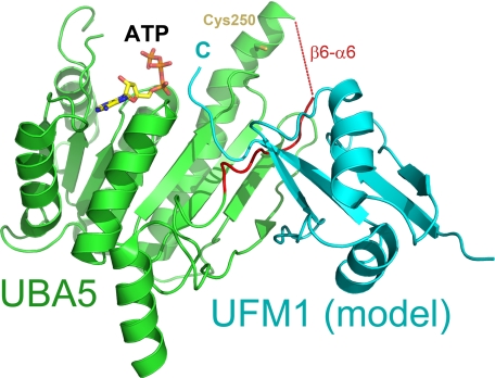
E1 ubiquitin-activating enzymes (UBAs) are large multidomain proteins that catalyze formation of a thioester bond between the terminal carboxylate of a ubiquitin or ubiquitin-like modifier (UBL) and a conserved cysteine in an E2 protein, producing reactive ubiquityl units for subsequent ligation to substrate lysines. Two important E1 reaction intermediates have been identified: a ubiquityl-adenylate phosphoester and a ubiquityl-enzyme thioester. However, the mechanism of thioester bond formation and its subsequent transfer to an E2 enzyme remains poorly understood. We have determined the crystal structure of the human UFM1 (ubiquitin-fold modifier 1) E1-activating enzyme UBA5, bound to ATP, revealing a structure that shares similarities with both large canonical E1 enzymes and smaller ancestral E1-like enzymes. In contrast to other E1 active site cysteines, which are in a variably sized domain that is separate and flexible relative to the adenylation domain, the catalytic cysteine of UBA5 (Cys(250)) is part of the adenylation domain in an alpha-helical motif. The novel position of the UBA5 catalytic cysteine and conformational changes associated with ATP binding provides insight into the possible mechanisms through which the ubiquityl-enzyme thioester is formed. These studies reveal structural features that further our understanding of the UBA5 enzyme reaction mechanism and provide insight into the evolution of ubiquitin activation.


Figures

FIGURE 1.
FIGURE 1.
UFM1 activation and loading activity of UBA5. 0.5 μm UBA5, 1 μm UFC1, and 1 μm UFM1 were incubated in a 10-μl reaction buffer containing 50 mm Tris-HCl, pH 7, 5 mm MgCl2, in the presence and absence of 1 μm ATP or 5 mm dithiothreitol at room temperature for 90 min. Products were resolved by nonreducing 10–20% gradient SDS-PAGE and immunoblotted with anti-UFM1 rabbit polyclonal primary (Boston Biochem) and anti-rabbit secondary antibodies (Thermo Scientific) followed by enhanced chemiluminescent detection (GE Healthcare). *, 5 mm dithiothreitol added to assay mix following initial reaction incubation. x, 10 μm UFC1 or UFM1. Full-length (Uba5l) and truncated (Uba5s) reaction products are indicated. C, C-terminal, N, N-terminal.
FIGURE 2.
FIGURE 2.
Ribbon representation of the UBA5 and related crystal structures. a, molecular surface representation of the UBA5 dimer. Catalytic cysteine is shown in stick format, and N and C termini are labeled. b, architecture of E1 and E1-like enzymes. Conserved adenylation domains (shown in blue) and SCCH domains (or structurally analogous regions; shown in orange) of the following are shown: MoeB (1JWA) (13), ThiF (1ZFN) (23), UBA5, UBA3 (1YOV) (28), UBA2 (1Y8Q) (15), UBA1 (3CMM) (30). Only one of the subunits of the heterodimeric E1s are shown. Distances between catalytic cysteines and A-site regions are shown with a black line. The flexible region in UBA5 between the crossover loop and the catalytic cysteine and the analogous flexible loop region in MoeB are indicated as straight light-orange lines. Catalytic cysteines are shown in stick format. All structure figures were prepared with PyMOL.
FIGURE 3.
FIGURE 3.
ATP binding pocket of UBA5. a, thermal stabilization by ATP. Solid and empty circles represent UBA5 protein in the presence and absence of 2 mm ATP, respectively. An increase in fluorescence is indicative of protein denaturation. Plots of fluorescence intensity versus temperature were fitted from the inflection point of the curves to interpolate the temperature at which 50% of the protein was unfolded. This transition temperature was increased by 3.2 °C in the presence of ATP. b, schematic diagram of hydrogen bonding network around ATP. ATP is shown in black, labeled side chains of UBA5 are shown in blue, and hydrogen bonds are shown as dashed lines. c, the ATP-binding active site of the A (green) and B (cyan) subunits are shown from the same perspective, side by side. Side chains that show structural variation in the two subunits and ATP are shown in stick format. Distances between Lys127 and ATP γ- and β-phosphates and ribose are 2.7, 3.5, and 2.7 Å, respectively. A. U., absorbance units.
FIGURE 4.
FIGURE 4.
Ribbon representations of the A and B subunits of UBA5. a, catalytic cysteine (Cys250), ATP, and boundaries of partially disordered loop regions (β2–α3 and β6–α6) are labeled. Helices, strands, and loops are colored cyan, green, and yellow, respectively. b, stereoscopic representation of the catalytic cysteine and its environs. Van der Waals interactions are shown as dashed black lines. The disordered region of the crossover loop is shown as a dashed blue line, and the hydrogen bond between the catalytic cysteine sulfur and the Asn210 main chain carbonyl is shown as a dashed red line. Secondary structures are labeled; B subunit labels are primed. Water molecules are shown as red stars. N, N-terminal; C, C-terminal.
FIGURE 5.
FIGURE 5.
Predicted model of the UBA5-UFM1 complex. UBA5 (green) and UFM1 (cyan) are shown as a schematic. The region encompassing the crossover loop is colored red. ATP and catalytic cysteine are shown in stick format and labeled. Position of the UFM1 C terminus (C) is also labeled.

References

    1. Schulman B. A., Harper J. W. (2009) Nat. Rev. Mol. Cell Biol. 10, 319–331 - PMC - PubMed
    1. Hochstrasser M. (2000) Nat Cell Biol. 2, E153–7 - PubMed
    1. Ciechanover A. (1998) EMBO J. 17, 7151–7160 - PMC - PubMed
    1. Schnell J. D., Hicke L. (2003) J. Biol. Chem. 278, 35857–35860 - PubMed
    1. Tang Z., Hecker C. M., Scheschonka A., Betz H. (2008) FEBS. J. 275, 3003–3015 - PubMed

Publication types

Associated data

LinkOut - more resources