

Structural determinants underlying photoprotection in the photoactive orange carotenoid protein of cyanobacteria
- PMID: 20368334
- PMCID: PMC2881762
- DOI: 10.1074/jbc.M110.115709
Structural determinants underlying photoprotection in the photoactive orange carotenoid protein of cyanobacteria
Abstract
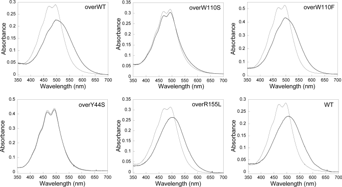
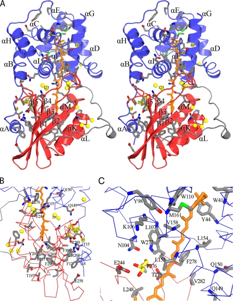
The photoprotective processes of photosynthetic organisms involve the dissipation of excess absorbed light energy as heat. Photoprotection in cyanobacteria is mechanistically distinct from that in plants; it involves the orange carotenoid protein (OCP), a water-soluble protein containing a single carotenoid. The OCP is a new member of the family of blue light-photoactive proteins; blue-green light triggers the OCP-mediated photoprotective response. Here we report structural and functional characterization of the wild type and two mutant forms of the OCP, from the model organism Synechocystis PCC6803. The structural analysis provides high resolution detail of the carotenoid-protein interactions that underlie the optical properties of the OCP, unique among carotenoid-proteins in binding a single pigment per polypeptide chain. Collectively, these data implicate several key amino acids in the function of the OCP and reveal that the photoconversion and photoprotective responses of the OCP to blue-green light can be decoupled.
Figures


Similar articles
-
A photoactive carotenoid protein acting as light intensity sensor.Proc Natl Acad Sci U S A. 2008 Aug 19;105(33):12075-80. doi: 10.1073/pnas.0804636105. Epub 2008 Aug 7. Proc Natl Acad Sci U S A. 2008. PMID: 18687902 Free PMC article.
-
Identification of a protein required for recovery of full antenna capacity in OCP-related photoprotective mechanism in cyanobacteria.Proc Natl Acad Sci U S A. 2010 Jun 22;107(25):11620-5. doi: 10.1073/pnas.1002912107. Epub 2010 Jun 7. Proc Natl Acad Sci U S A. 2010. PMID: 20534537 Free PMC article.
-
In vitro reconstitution of the cyanobacterial photoprotective mechanism mediated by the Orange Carotenoid Protein in Synechocystis PCC 6803.Plant Cell. 2011 Jul;23(7):2631-43. doi: 10.1105/tpc.111.086884. Epub 2011 Jul 15. Plant Cell. 2011. PMID: 21764991 Free PMC article.
-
The Orange Carotenoid Protein: a blue-green light photoactive protein.Photochem Photobiol Sci. 2013 Jul;12(7):1135-43. doi: 10.1039/c3pp25406b. Photochem Photobiol Sci. 2013. PMID: 23396391 Review.
-
Structure and functions of Orange Carotenoid Protein homologs in cyanobacteria.Curr Opin Plant Biol. 2017 Jun;37:1-9. doi: 10.1016/j.pbi.2017.03.010. Epub 2017 Apr 6. Curr Opin Plant Biol. 2017. PMID: 28391046 Review.
Cited by
-
A comparative study of three signaling forms of the orange carotenoid protein.Photosynth Res. 2016 Dec;130(1-3):389-401. doi: 10.1007/s11120-016-0272-8. Epub 2016 May 9. Photosynth Res. 2016. PMID: 27161566
-
Single-molecule trapping and spectroscopy reveals photophysical heterogeneity of phycobilisomes quenched by Orange Carotenoid Protein.Nat Commun. 2019 Mar 12;10(1):1172. doi: 10.1038/s41467-019-09084-2. Nat Commun. 2019. PMID: 30862823 Free PMC article.
-
The photocycle of orange carotenoid protein conceals distinct intermediates and asynchronous changes in the carotenoid and protein components.Sci Rep. 2017 Nov 14;7(1):15548. doi: 10.1038/s41598-017-15520-4. Sci Rep. 2017. PMID: 29138423 Free PMC article.
-
Engineering hydrogen bonding at tyrosine-201 in the orange carotenoid protein using halogenated analogues.Photosynth Res. 2025 Jan 20;163(1):10. doi: 10.1007/s11120-024-01133-2. Photosynth Res. 2025. PMID: 39832061
-
Structural and functional modularity of the orange carotenoid protein: distinct roles for the N- and C-terminal domains in cyanobacterial photoprotection.Plant Cell. 2014 Jan;26(1):426-37. doi: 10.1105/tpc.113.118588. Epub 2014 Jan 7. Plant Cell. 2014. PMID: 24399299 Free PMC article.
References
-
- Demmig-Adams B. (1990) Biochim. Biophys. Acta 1020, 1–24
-
- Horton P., Ruban A. V., Walters R. G. (1996) Annu. Rev. Plant Physiol. Plant Mol. Biol. 47, 655–684 - PubMed
-
- Niyogi K. K. (1999) Annu. Rev. Plant Physiol. Plant Mol. Biol. 50, 333–359 - PubMed
-
- Gilmore A. M., Yamamoto H. Y. (1993) Photosynth. Res. 35, 67–78 - PubMed
Publication types
MeSH terms
Substances
Associated data
- Actions
- Actions
- Actions
LinkOut - more resources
Full Text Sources
Other Literature Sources
Molecular Biology Databases
