. 2010 Jun;78(6):2584-98.
doi: 10.1128/IAI.00141-10. Epub 2010 Apr 5.

The C-terminal tail of Yersinia pseudotuberculosis YopM is critical for interacting with RSK1 and for virulence

Affiliations

The C-terminal tail of Yersinia pseudotuberculosis YopM is critical for interacting with RSK1 and for virulence

Melissa W McCoy et al. Infect Immun. 2010 Jun.

Abstract

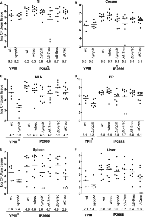
Yersinia spp. undermine the immune responses of infected animals by translocating Yops directly into host cells with a type III secretion system. YopM, a leucine-rich repeat protein, is a critical virulence factor in infection. YopM localizes to both the nucleus and the cytoplasm in cultured cells, interacts with mammalian p90 ribosomal S6 kinase 1 (RSK1), and causes a decrease in NK cell populations in spleens. Little is known about the molecular interaction between YopM and RSK1 and its significance in pathogenesis. We performed a systematic deletion analysis of YopM in Yersinia pseudotuberculosis to determine which regions are required for RSK1 interactions, nuclear localization, virulence, and changes in immune cell populations during infection of mice. Full-length YopM associated with RSK1 in at least two protein complexes in infected cells, and deletion of its C-terminal tail abrogated all RSK1 interactions. The C-terminal tail was required for tissue colonization, as yopM mutants that failed to interact with RSK1 were as defective for tissue colonization as was a DeltayopM mutant; however, nuclear localization of YopM was not dependent on its RSK1 interaction. Mutants expressing YopM proteins which do not interact with RSK1 caused more pathology than did the DeltayopM mutant, suggesting that there are other RSK1-independent functions of YopM. Histopathological and flow cytometric analyses of spleens showed that infection with wild-type Y. pseudotuberculosis caused an influx of neutrophils, while mice infected with yopM mutants had increased numbers of macrophages. Decreases in NK cells after Y. pseudotuberculosis infection did not correlate with YopM expression. In conclusion, the C terminus of YopM is essential for RSK1 interactions and for virulence.


Figures

FIG. 1.
FIG. 1.
YopM-RSK1 complex formation requires the C-terminal tail of YopM. (A) Schematic of YopM protein and location of LRRs 1 to 15. Regions deleted in each LRR mutant are indicted below the first and last amino acids deleted in each mutant. Each deleted region was replaced with a single glycine residue. (B to D) RAW 264.7 macrophages were infected for 30 min with strains of YPIIIΔyopM harboring vector alone or plasmids expressing full-length YopM, LRR deletion mutants, or ΔC or with YPIIIΔyscB--yscL to control for bacterial lysis. Mammalian cells were lysed, and proteins were separated by denaturing (B) or nondenaturing (C and D) PAGE. Protein expression and complex formation were analyzed by immunoblot analysis, using antibodies against YopM (B and C), RSK1 (D), or β-actin (B). The lane labeled YopMpur (B) is from a shorter exposure of the film and contains YopM protein purified from E. coli. Macrophages infected with YPIIIΔyopM plus Δ8-9 were processed on a different nondenaturing gel. The masses of native molecular weight markers in kilodaltons are indicated on the left of the native blots. YMC1 and YMC2 are indicated on the right of panels C and D. These assays were repeated at least six times.
FIG. 2.
FIG. 2.
The last six amino acids of YopM are required for interaction with RSK1. (A) Schematic of the eight sets of triple alanine substitutions engineered into the last 24 amino acids of YopM, called C1 to C8. (B to D) RAW 264.7 macrophages were infected for 30 min with strains of YPIIIΔyopM harboring vector alone or plasmids expressing full-length YopM or the indicated mutants or with YPIIIΔyscB-yscL to control for bacterial lysis. Mammalian cells were lysed, and proteins were separated by denaturing (B) or nondenaturing (C and D) PAGE. Protein expression and complex formation were analyzed by immunoblot analysis, using anti-YopM (B and C) and anti-RSK1 (D) antibodies. Numbers on the left of the blots indicate the masses of molecular weight markers in kilodaltons. These assays were repeated at least two times.
FIG. 3.
FIG. 3.
YopM and ΔC form multimers in solution. YopM and ΔC proteins were purified after overexpression in E. coli, separated by nondenaturing PAGE, and visualized by staining with Coomassie brilliant blue. Masses of native molecular weight markers in kilodaltons are indicated on the left.
FIG. 4.
FIG. 4.
The C terminus of YopM is not required for nuclear localization. RAW 264.7 cells were infected for 30 min with strains of YPIIIΔyopM harboring vector alone or plasmids expressing full-length YopM or the indicated mutants or with YPIIIΔyscB-yscL to control for bacterial lysis. Cytoplasmic (C) and nuclear (N) fractions of the infected cells were prepared. Proteins were separated by SDS-PAGE and subjected to Western blot analysis. Blots were analyzed with antibodies against YopM (A) and the cytoplasmic protein Rho GDI (C), and then the YopM blot was stripped and reprobed with antibodies to the nuclear protein CREB (B). Numbers on the left side of the blots indicate masses of molecular weight markers in kilodaltons. This assay was repeated at least four times.
FIG. 5.
FIG. 5.
YopM-RSK1 interactions required for YMC1 are critical for colonization of spleen and lungs in intravenously infected mice. (A) Translocation assay of YopM proteins expressed by IP2666 WTrec and yopM mutant strains used to infect mice. YPIIIΔyscB-yscL was included to control for bacterial lysis. (B to D) Seven- to 8-week-old BALB/c mice were infected intravenously with 100 CFU of IP2666 WTrec or yopM mutant strains. Four days postinfection, mice were sacrificed and the numbers of bacteria in the spleen (B), lung (C), and liver (D) were determined. Closed circles represent the log10 CFU/gram of tissue from an individual mouse, and open circles indicate that the CFU were below the limit of detection. Bars represent the geometric means, which are also written below each strain as their log10 value. P values were determined using ANOVA followed by Dunnett's multiple-comparison test. Single asterisks indicate statistical significance of P < 0.05 between WTrec and a mutant strain.
FIG. 6.
FIG. 6.
Colonization of YopM mutants after orogastric infection of mice. Seven- to 8-week-old female BALB/c mice were orogastrically infected with either 2 × 10⁹ (YPIII) or 7 × 10⁸ (IP2666) CFU of WT, WTrec, or mutant strains. Four days post-infection with IP2666 or 5 days post-infection with YPIII strains, mice were sacrificed and the numbers of Y. pseudotuberculosis bacteria in the small intestine (SI), cecum, Peyer's patches (PP), mesenteric lymph nodes (MLN), spleen, and liver were determined. Closed circles represent the log10 CFU/gram of tissue from one mouse, open circles indicate that the CFU were below the limit of detection, and bars represent the geometric means, which are also written below each strain as their log10 value. Statistical significance of P < 0.05 between the WT and the ΔyopM mutant for the YPIII strains was determined by t test and is indicated by a single asterisk. Statistical significance of P < 0.05 between WT IP2666 and other IP2666 strains was determined by ANOVA followed by Dunnett's multiple-comparison test.
FIG. 7.
FIG. 7.
Interaction with RSK1 is not responsible for all pathological effects of YopM. Spleens from mice mock infected with PBS (A) or infected with IP2666 WTrec (B), ΔyopM (C), C7rec (D), or C8rec (E) strains for 4 days were subjected to histopathological analysis by staining with hematoxylin and eosin. Each micrograph shows an example of the pathology caused by each strain. Pathologies are marked as follows: inflammation (formula image), necrosis (formula image), and congestion (formula image). Scale bars represent 100 μm.
FIG. 8.
FIG. 8.
YopM increases the numbers of neutrophils while the ΔyopM mutation increases the numbers of macrophages in infected spleens. Splenocytes were prepared from mice mock infected intravenously with PBS or infected with the IP2666 WTrec, ΔyopM, C7rec, or C8rec strain. Four days postinfection, percentages of splenocytes that were macrophages and neutrophils/inflammatory monocytes were determined by flow cytometry using PE-CD11b and FITC-GR1 antibodies. (A) Graphic plot of macrophages and neutrophils/inflammatory monocytes (GR1high cells) from a mock-infected spleen sample, as measured by levels of staining for the surface markers CD11b and GR1. Numbers next to gates indicate percentages of splenocytes in each gate. (B) Percentages of splenocytes in mice infected with each strain that were GR1high cells. (C) Percentages of splenocytes in infected mice that were macrophages. Each circle represents the data from one mouse, and bars represent the means. P values were determined using ANOVA followed by Dunnett's multiple-comparison test. Single asterisks indicate statistical significance of P < 0.05 between mock-infected cells and cells infected with the indicated strain.
FIG. 9.
FIG. 9.
CD49b+ NK cells are reduced in a YopM-independent manner. (A) Graphic plot of NK cells from a mock-infected spleen sample, as measured by levels of staining for the surface markers CD49b and CD3. Numbers next to gates indicate percentages of splenocytes in gates. (B) Percentages of splenocytes that were NK cells in mock-infected mice and mice infected with the WTrec, ΔyopM, C7rec, or C8rec strain. Each circle represents the data from one mouse, and bars represent the means. A single asterisk indicates significant differences between NK cell levels in mock-infected mice and NK cell levels in Yersinia-infected mice as determined by ANOVA followed by Dunnett's multiple-comparison posttest. Double asterisks indicate significant differences between the levels of NK cells in WTrec-infected mice and those in mice infected with the ΔyopM, C7rec, or C8rec strain and are shown in the second row beneath the strains. (C and D) Percentages of splenocytes that were NK cells, as a function of CFU/spleen, in mice infected with various doses of the ΔyopM strain (C) and either the ΔyopE or ΔyopH strain (D). Correlations were significant with values of P = 0.039 (ΔyopM) and P < 0.01 (ΔyopEyopH), Pearson r values of −0.554 (ΔyopM) and −0.697 (ΔyopE,ΔyopH), and R2 values of 0.307 (ΔyopM) and 0.75 (ΔyopE,ΔyopH). (E) Fold increase, compared to mock-infected mice, in CD69 expression on CD49bhigh NK cells as a function of the fold decrease in NK cells. Correlation was significant with P < 0.0001, a Pearson r value of 0.868, and an R2 value of 0.75. For panels C to E, each circle represents the data from one mouse. MFI, mean fluorescence intensity.

References

    1. Arase, H., T. Saito, J. H. Phillips, and L. L. Lanier. 2001. Cutting edge: the mouse NK cell-associated antigen recognized by DX5 monoclonal antibody is CD49b (alpha 2 integrin, very late antigen-2). J. Immunol. 167:1141-1144. - PubMed
    1. Balada-Llasat, J. M., and J. Mecsas. 2006. Yersinia has a tropism for B and T cell zones of lymph nodes that is independent of the type III secretion system. PLoS Pathog. 2:e86. - PMC - PubMed
    1. Barnes, P. D., M. A. Bergman, J. Mecsas, and R. R. Isberg. 2006. Yersinia pseudotuberculosis disseminates directly from a replicating bacterial pool in the intestine. J. Exp. Med. 203:1591-1601. - PMC - PubMed
    1. Bella, J., K. L. Hindle, P. A. McEwan, and S. C. Lovell. 2008. The leucine-rich repeat structure. Cell. Mol. Life Sci. 65:2307-2333. - PMC - PubMed
    1. Benabdillah, R., L. J. Mota, S. Lutzelschwab, E. Demoinet, and G. R. Cornelis. 2004. Identification of a nuclear targeting signal in YopM from Yersinia spp. Microb. Pathog. 36:247-261. - PubMed

Publication types

MeSH terms

Substances

LinkOut - more resources