. 2010 Jun;78(6):2700-13.
doi: 10.1128/IAI.01389-09. Epub 2010 Apr 5.

Salmonella enterica serovar typhimurium invades fibroblasts by multiple routes differing from the entry into epithelial cells

Affiliations

Salmonella enterica serovar typhimurium invades fibroblasts by multiple routes differing from the entry into epithelial cells

Ana Aiastui et al. Infect Immun. 2010 Jun.

Abstract

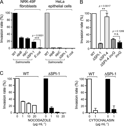
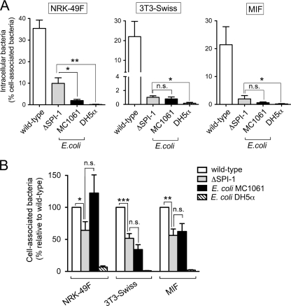
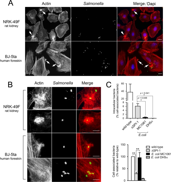
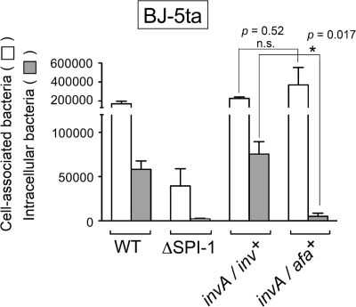
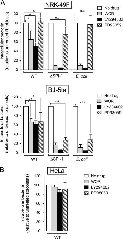
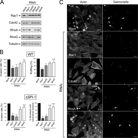
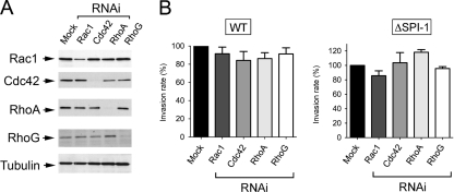
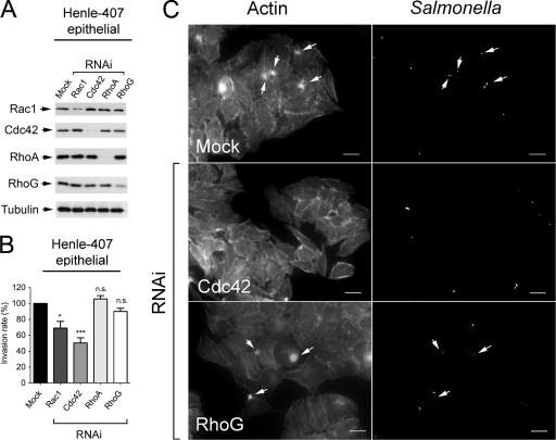
Fibroblasts are ubiquitous cells essential to tissue homeostasis. Despite their nonphagocytic nature, fibroblasts restrain replication of intracellular bacterial pathogens such as Salmonella enterica serovar Typhimurium. The extent to which the entry route of the pathogen determines this intracellular response is unknown. Here, we analyzed S. Typhimurium invasion in fibroblasts obtained from diverse origins, including primary cultures and stable nontransformed cell lines derived from normal tissues. Features distinct to the invasion of epithelial cells were found in all fibroblasts tested. In some fibroblasts, bacteria lacking the type III secretion system encoded in the Salmonella pathogenicity island 1 displayed significant invasion rates and induced the formation of lamellipodia and filopodia at the fibroblast-bacteria contact site. Other bacterial invasion traits observed in fibroblasts were the requirement of phosphatidylinositol 3-kinase, mitogen-activated protein kinase MEK1, and both actin filaments and microtubules. RNA interference studies showed that different Rho family GTPases are targeted by S. Typhimurium to enter into distinct fibroblasts. Rac1 and Cdc42 knockdown affected invasion of normal rat kidney fibroblasts, whereas none of the GTPases tested (Rac1, Cdc42, RhoA, or RhoG) was essential for invasion of immortalized human foreskin fibroblasts. Collectively, these data reveal a marked diversity in the modes used by S. Typhimurium to enter into fibroblasts.


Figures

FIG. 1.
FIG. 1.
Salmonella invasion of NRK-49F fibroblasts show features distinct to the invasion of epithelial cells. (A) Invasion rates estimated by gentamicin survival assays and made relative to the rate of the wild-type bacteria. Incubation time with bacteria was 20 min. Viable intracellular bacteria were enumerated at 2 h postinfection and accounted, in the case of wild-type (WT) bacteria, for 1.04% and 3.60% of the inoculum in NRK-49F fibroblasts and HeLa epithelial cells, respectively. The S. Typhimurium strains used were SL1344 (wild-type), SB169 (sipB), SB220 (sipC), and MD706 (ΔSPI-1). The E. coli motile strain MC1061 was included for comparison. (B) Dispensability of the type I secretion system encoded by SPI-4 for bacterial entry into NRK-49F fibroblasts. The S. Typhimurium strains used included MD173 (invG), MvP602 (ΔSPI-4), and MvP603 (ΔSPI-4 invG). (C) Inhibitory effect of nocodazole and cytochalasin on the invasion of NRK-49F fibroblasts by S. Typhimurium wild-type and ΔSPI-1 strains. The invasion rate of the ΔSPI-1 strain (16% of that of wild-type strain) was normalized to 100% to highlight the differences in the drug-treated fibroblasts. Data are the means and standard deviations from three independent experiments.
FIG. 2.
FIG. 2.
S. Typhimurium entry into NRK-49F fibroblasts occurs via two modes that differ in the extent of actin rearrangement and macropinocytosis. Extracellular (blue) and intracellular (red) bacteria were differentiated by the in-out staining procedure (see Materials and Methods). Actin was labeled with fluorescein isothiocyanate-phalloidin (green). Entry of S. Typhimurium wild-type (WT) and ΔSPI-1 bacteria was compared to that of the E. coli MC1061 motile strain. (A) Low-magnification images showing prominent membrane ruffling and macropinocytosis induced by S. Typhimurium wild-type bacteria in NRK-49F fibroblasts. Note that, unlike the few E. coli MC1061 cells observed associated to the fibroblasts, some of the S. Typhimurium ΔSPI-1 bacteria were associated to lamellipodium-like membrane extensions. Bar, 10 μm. (B) Magnifications of the areas marked in panel A. Arrows indicate a large and actin-rich lamellipodium contacting a surface-located ΔSPI-1 bacterium and discrete actin accumulations surrounding two E. coli MC1061 bacteria located intracellularly. Bar, 5 μm. (C) Scanning electron microscopy images of NRK-49F fibroblasts infected for 20 min with S. Typhimurium wild-type or ΔSPI-1 bacteria. Note the prominent membrane ruffling in the area where wild-type bacteria locate. Filopodia and lamellipodia were observed associated to ΔSPI-1 bacteria located onto the fibroblast surfaces. The ΔSPI-1 mutant was also occasionally visualized retracting the fibroblast surface underneath invading bacteria (arrows).
FIG. 3.
FIG. 3.
Requirement of the TTSS encoded by SPI-1 for entry into diverse fibroblast cells. The cell lines used included NRK-49F, 3T3-Swiss fibroblasts, and MIF. (A) Invasion rates of S. Typhimurium SL1344 (wild type) and MD706 (ΔSPI-1) and the E. coli strains MC1061 (motile) and DH5α (nonmotile) expressed as the ratio of intracellular bacteria versus total number of fibroblast-associated bacteria counted before gentamicin treatment. Using this parameter, the invasiveness of the ΔSPI-1 mutant relative to the wild type was 28% (NRK-49F), 4.6% (3T3-Swiss fibroblasts), and 9.2% (MIF). (B) Bacterial adhesion to the different fibroblast cell lines used. The number of cell-associated bacteria was made relative to that of wild-type strain in each of the fibroblast cell lines used. Data are the means and standard deviations from three independent experiments. *, P = 0.01 to 0.05; **, P = 0.001 to 0.01; n.s., not significant by a Student t test (A) and one-way ANOVA with Tukey's posttest (B).
FIG. 4.
FIG. 4.
The extent of membrane ruffling induced by S. Typhimurium in fibroblasts varies depending on the source of fibroblasts used. (A) Actin distribution in NRK-49F fibroblasts and BJ-5ta fibroblasts infected with the S. Typhimurium SL1344 (wild-type) strain for 20 min. Note that the ruffles are more prominent in the infected NRK-49F fibroblasts. Actin was labeled with Alexa-Fluor 594-phalloidin (red), bacteria was labeled with primary anti-rabbit Salmonella followed by secondary Alexa-Fluor 488-conjugated goat anti-rabbit antibody (green), and the nuclei were labeled with DAPI (blue). Bar, 10 μm. (B) Enlargement of areas marked with arrows in panel A. Bar, 5 μm. (C) Invasion rates and adherence properties of the S. Typhimurium ΔSPI-1 mutant and the E. coli strains MC1061 and DH5α in the BJ-5ta fibroblasts. Data are the means and standard deviations from three independent experiments. *, P = 0.01 to 0.05; **, P = 0.001 to 0.01; n.s., not significant (Student t test).
FIG. 5.
FIG. 5.
Increased adherence to BJ-5ta fibroblasts mediated by the afimbrial adhesin AFA-I is not followed by a higher invasion rate in the SPI-1-independent entry mode. Shown are absolute numbers of fibroblast-associated bacteria counted before gentamicin treatment and those of intracellular bacteria surviving gentamicin treatment. Infection was for 20 min (wild type) or 60 min (all other strains). The S. Typhimurium strains used included SL1344 (WT), MD706 (ΔSPI-1), MD1662 (invA/inv+) and MD1663 (invA/afa+). Note that the expression of the invasin protein (Inv) from Y. pseudotuberculosis increases bacterial entry while the enhanced adherence promoted by the afimbrial adhesin AFA-I (compared to the ΔSPI-1 strain) does not. Data are the means and standard deviations from three independent experiments. *, P = 0.01 to 0.05; n.s., not significant (Student t test).
FIG. 6.
FIG. 6.
PI3K and the MAP kinase MEK1 are involved to different extents in the entry into fibroblasts of S. Typhimurium SL1344 (wild-type) and MD706 (ΔSPI-1) strains and the E. coli motile strain MC1061. The role of these two kinases was assessed in invasion assays using 100 nM wortmannin (WOR) and 25 μM LY294002 (PI3K inhibitors) or 50 μM PD98059 (MEK1 inhibitor). (A) Effect of the drugs on bacterial entry into NRK-49F fibroblasts and BJ-5ta fibroblasts. (B) Control assay performed in HeLa human epithelial cells. Note the dispensability of these two kinases in HeLa epithelial cells and their differential requirement for bacterial entry into fibroblasts depending on the fibroblast and the bacterial strain used. Data are the means and the standard deviations of three independent experiments. *, P = 0.01 to 0.05; **, P = 0.001 to 0.01; ***, P < 0.001; n.s., not significant (Student t test).
FIG. 7.
FIG. 7.
Requirement of Rho family GTPases for entry of S. Typhimurium SL1344 (wild type) and MD706 (ΔSPI-1) strains into NRK-49F fibroblasts. (A) Levels of Rac1, Cdc42, RhoA, and RhoG upon incubation for 72 h in the presence of their respective RNAi. The level of α-tubulin was determined as a control. (B) Invasion rate of wild-type (WT) and ΔSPI-1 bacteria in RNAi-treated NRK-49F fibroblasts. Incubation time with bacteria was of 20 min (wild type) and 40 min (ΔSPI-1). Viable intracellular bacteria were counted at 2 h postinfection. Shown are the percentages of RNAi-treated NRK-49F fibroblasts either exhibiting membrane ruffling in response to the infection with wild-type bacteria or containing intracellular bacteria upon infection with the ΔSPI-1 mutant. The analysis in the ΔSPI-1 mutant was performed with in-out staining. Data are the means and the standard deviations of three independent experiments. *, P = 0.01 to 0.05; **, P = 0.001 to 0.01; ***, P < 0.001; n.s., not significant (one-way ANOVA with Tukey's posttest). (C) Microscopy analysis showing the capacity of wild-type bacteria to induce membrane ruffling in NRK-49F fibroblasts depleted of RhoA or RhoG. Bar, 10 μm.
FIG. 8.
FIG. 8.
RNAi interference directed at Rac1, Cdc42, RhoA, or RhoG does not substantially affect the invasion of BJ-5ta fibroblasts by S. Typhimurium SL1344 (wild type) or MD706 (ΔSPI-1) strains. (A) Levels of Rac1, Cdc42, RhoA, and RhoG upon incubation for 72 h in the presence of their respective RNAi. The level of α-tubulin was determined as a control. (B) Invasion rate of wild-type (WT) and ΔSPI-1 bacteria in RNAi-treated BJ-5ta fibroblasts. Incubation times with bacteria were 20 min (wild type) and 60 min (ΔSPI-1). Viable intracellular bacteria were counted at 2 h postinfection. Data were analyzed by one-way ANOVA with Tukey's posttest. Shown are the means and the standard deviations of three independent experiments. No significant differences were found.
FIG. 9.
FIG. 9.
Rac1 and Cdc42 are targeted by S. Typhimurium SL1344 (wild type) to invade Henle-407 human epithelial cells. (A) Levels of Rac1, Cdc42, RhoA, and RhoG upon incubation for 72 h in the presence of their respective RNAi. The level of α-tubulin was determined as a control. (B) Invasion rate of wild-type bacteria in RNAi-treated Henle-407 human epithelial cells. Incubation time with bacteria was 20 min, and viable intracellular bacteria were counted at 2 h postinfection. Data are the means and the standard deviations of three independent experiments. *, P = 0.01 to 0.05; ***, P < 0.001; n.s., not significant (one-way ANOVA with Tukey's posttest). Note that although Rac1 and RhoG are not totally depleted by the RNAi treatment, a significant effect on invasion is observed in the case of only Rac1. (C) Representative microscopy images depicting the involvement of Cdc42 in the membrane ruffling triggered by wild-type bacteria in these epithelial cells. Bar, 10 μm.

References

    1. Aspenstrom, P., A. Fransson, and J. Saras. 2004. Rho GTPases have diverse effects on the organization of the actin filament system. Biochem. J. 377:327-337. - PMC - PubMed
    1. Bakowski, M. A., V. Braun, and J. H. Brumell. 2008. Salmonella-containing vacuoles: directing traffic and nesting to grow. Traffic 9:2022-2031. - PubMed
    1. Baumler, A. J., R. M. Tsolis, and F. Heffron. 1996. Contribution of fimbrial operons to attachment to and invasion of epithelial cell lines by Salmonella typhimurium. Infect. Immun. 64:1862-1865. - PMC - PubMed
    1. Bishop, A., D. House, T. Perkins, S. Baker, R. A. Kingsley, and G. Dougan. 2008. Interaction of Salmonella enterica serovar Typhi with cultured epithelial cells: roles of surface structures in adhesion and invasion. Microbiology 154:1914-1926. - PMC - PubMed
    1. Biswas, D., K. Itoh, and C. Sasakawa. 2003. Role of microfilaments and microtubules in the invasion of INT-407 cells by Campylobacter jejuni. Microbiol. Immunol. 47:469-473. - PubMed

Publication types

Substances