. 2010 Jun;30(11):2608-20.
doi: 10.1128/MCB.00208-09. Epub 2010 Apr 5.

The Ras effector RASSF2 controls the PAR-4 tumor suppressor

Affiliations

The Ras effector RASSF2 controls the PAR-4 tumor suppressor

Howard Donninger et al. Mol Cell Biol. 2010 Jun.

Abstract

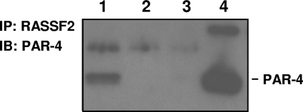
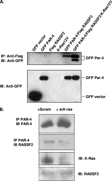
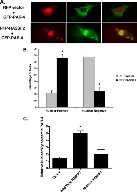
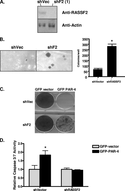
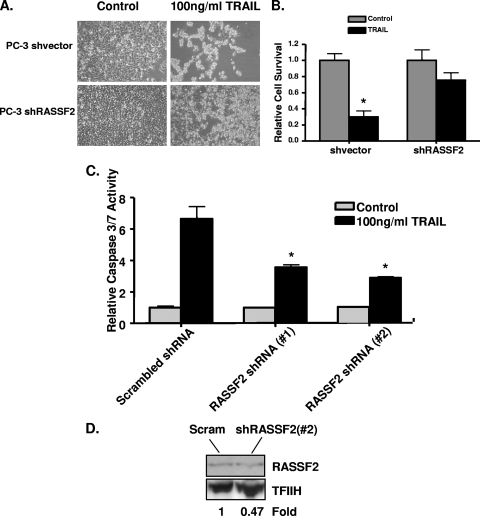
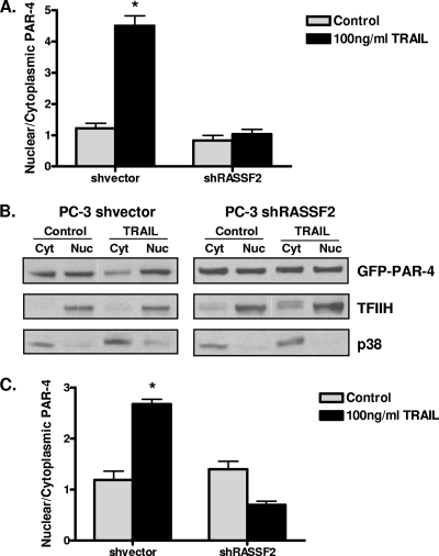
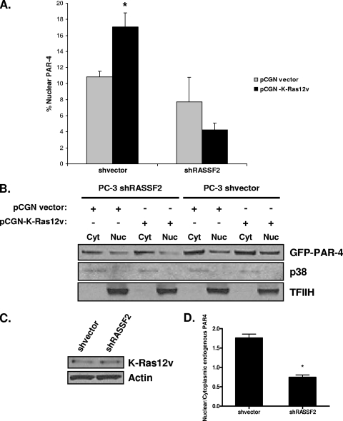
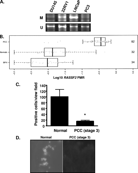
RASSF2 is a novel proapoptotic effector of K-Ras. Inhibition of RASSF2 expression enhances the transforming effects of K-Ras, and epigenetic inactivation of RASSF2 is frequently detected in mutant Ras-containing primary tumors. Thus, RASSF2 is implicated as a tumor suppressor whose inactivation facilitates transformation by disconnecting apoptotic responses from Ras. The mechanism of action of RASSF2 is not known. Here we show that RASSF2 forms a direct and endogenous complex with the prostate apoptosis response protein 4 (PAR-4) tumor suppressor. This interaction is regulated by K-Ras and is essential for the full apoptotic effects of PAR-4. RASSF2 is primarily a nuclear protein, and shuttling of PAR-4 from the cytoplasm to the nucleus is essential for its function. We show that RASSF2 modulates the nuclear translocation of PAR-4 in prostate tumor cells, providing a mechanism for its biological effects. Thus, we identify the first tumor suppressor signaling pathway emanating from RASSF2, we identify a novel mode of action of a RASSF protein, and we provide an explanation for the extraordinarily high frequency of RASSF2 inactivation we have observed in primary prostate tumors.


Figures

FIG. 1.
FIG. 1.
RASSF2 interacts with PAR-4 at physiologically relevant levels. Lysates with equal protein concentrations from H441 lung cancer cells were immunoprecipitated with an anti-RASSF2 antibody (lane 1) or control IgG (lane 2), fractionated on SDS gels, and immunoblotted (IB) with an anti-PAR-4 antibody. Lane 3 is a mock IP of a buffer with no lysate. The endogenous interaction between RASSF2 and PAR-4 was confirmed by the presence of PAR-4 in the proteins precipitated with the RASSF2 antibody (lane 1) and not with control IgG (lane 2). Lane 4 is a PAR-4 positive control lysate.
FIG. 2.
FIG. 2.
Activated K-Ras enhances the interaction between RASSF2 and PAR-4. (A) 293T cells were transfected with Flag-RASSF2 and GFP-PAR-4 or GFP-vector in the presence or absence of activated K-Ras, and lysates were prepared and immunoprecipitated with anti-Flag, fractionated on SDS gels, and immunoblotted (IB) with anti-GFP (top panel). Activated K-Ras enhanced the association between RASSF2 and PAR-4 (compare lanes 5 and 6). Aliquots of the input lysates were similarly blotted with anti-GFP to ensure equivalent GFP-PAR-4 expression levels (bottom panel). The lower band in lane 6, top panel, was consistently observed and may correspond to a cleaved form of PAR-4. (B) H441 cells were transfected with K-ras siRNA or scrambled control. The cells were then lysed and immunoprecipitated with anti-PAR-4 antibodies. The immunoprecipitate was then Western blotted for endogenous RASSF2. The top panel shows that the siRNA did not inhibit the expression of PAR-4. The second panel shows that the levels of RASSF2 coprecipitating with PAR-4 were reduced in the siRNA K-Ras-treated cells. The third panel shows that K-Ras protein was downregulated in the siRNA treated cells. The fourth panel shows that the total levels of RASSF2 in the pre-IP lysates were the same.
FIG. 3.
FIG. 3.
RASSF2 promotes the nuclear localization of PAR-4. (A) COS-7 cells were cotransfected with RFP vector or RFP-RASSF2 and GFP-PAR-4, and images were captured 24 h later using a fluorescence microscope as described in Materials and Methods. In the absence of RASSF2, PAR-4 localized to the plasma membrane and cytoplasm, but in the presence of RASSF2, PAR-4 colocalized with RASSF2 in the nucleus, as evidenced by the yellow color in the merged image. Magnification (all images), ×100. (B) Quantification of PAR-4 nuclear localization in the presence of RASSF2. Fifty randomly selected cells expressing both RFP-RASSF2 or RFP-vector and GFP-PAR-4 were scored for the presence of nuclear PAR-4. The bars show the means of triplicate experiments, and standard deviations are indicated. *, statistically significantly different (P < 0.01) from vector-transfected cells. (C) PC-3 human prostate tumor cells were transfected with RASSF2 or a deletion mutant of RASSF2 lacking at least one of the NLSs (MutNLS) and fractionated before Western blotting for endogenous PAR-4. The blots were quantified and used to generate a ratio of nuclear versus cytoplasmic PAR-4. The mutant of RASSF2 was impaired for inducing the nuclear localization of endogenous PAR-4. *, statistically different (P < 0.01) from vector-transfected cells.
FIG. 4.
FIG. 4.
Loss of RASSF2 enhances tumorigenicity of prostate cancer cells and confers resistance to PAR-4-mediated cell death. (A) Western blot analysis of RASSF2 expression in PC-3 prostate cancer cells stably expressing a RASSF2 shRNA construct or corresponding vector control. Actin was used as a control for protein loading. (B) PC-3 cells stably expressing an shRNA to RASSF2 or vector control were plated in soft agar and scored for growth after 14 days. Quantification is shown in the adjacent panel. *, statistically different (P < 0.05) from cells transfected with the vector control. (C and D) The PC-3 prostate cancer cells stably expressing an shRNA to RASSF2 or a vector control were transfected with GFP-vector or GFP-PAR-4, and surviving colonies stained with crystal violet after 2 weeks of selection in G418 (C) or assayed for apoptosis via determination of caspase-3 and -7 activities (D). *, significantly different (P < 0.05) from cells transfected with GFP-vector.
FIG. 5.
FIG. 5.
Loss of RASSF2 protects prostate cancer cells from TRAIL-induced apoptosis. (A and B) PC-3 cells stably transfected with a RASSF2 shRNA construct or a control vector were seeded at 1 × 10⁵cells/well in six-well plates and treated with 100 ng/ml TRAIL, and cell death was estimated 72 h later by trypan blue exclusion. Bars show the means of triplicate experiments, and standard deviations are indicated. *, P < 0.05 compared to control cells. (C) Similar experiments were then performed to measure apoptosis. These included a second RASSF2 shRNA cell line (# 2). Cells were seeded at 3 × 10³ cells/well in 96-well plates, and caspase-3 and -7 activities were measured after TRAIL treatment. Caspase activity is expressed relative to results in untreated cells. The bars show means of duplicate experiments. with standard deviations shown. *, significantly different (P < 0.05) from cells transfected with a scrambled shRNA. (D) Western blot analysis of RASSF2 expression in PC-3 prostate cancer cells stably expressing a RASSF2 shRNA (#2) construct (#2) or scrambled shRNA. TFIIH was used as a loading control.
FIG. 6.
FIG. 6.
Loss of RASSF2 impairs TRAIL-induced PAR-4 nuclear trafficking in prostate cancer cells. PC-3 cells stably expressing a RASSF2 shRNA construct or control vector were transfected with GFP-PAR-4 and treated with 100 ng/ml TRAIL for 1 h. (A) Cells were lysed, and nuclear (Nuc) and cytoplasmic (Cyt) fractions were prepared and analyzed by Western blotting for GFP-PAR-4. Densitometric quantitation of Western blot results is shown. Bars show means of triplicate experiments, with standard deviations indicated. *, statistically different (P < 0.01) from untreated cells. (B) Representative Western blot. TFIIH and p38 were used as markers for the nuclear and cytoplasmic fractions, respectively. Similar experiments were performed for the endogenous PAR-4 protein. (C) Densitometric quantitation of the Western blot results with endogenous PAR-4 in nuclear and cytoplasmic fractions in the RASSF2 (positive and negative) PC-3 cells. Bars show means of triplicate experiments, and standard deviations are indicated. *, P < 0.01 compared to untreated cells.
FIG. 7.
FIG. 7.
Loss of RASSF2 reduces activated K-Ras-induced PAR-4 nuclear translocation. (A) PC-3 cells stably knocked down for RASSF2 and control cells were transfected with GFP-PAR-4 in the presence or absence of activated K-Ras. At 16 h posttransfection, cells were lysed and nuclear (Nuc) and cytoplasmic (Cyt) fractions were prepared and analyzed by Western blotting for GFP-PAR-4. The percentage of nuclear PAR-4 was quantitated by densitometry. Bars show means of duplicate experiments, and standard deviations are indicated. *, P < 0.05 compared to cells transfected with pCGN-vector. (B) Representative Western blots of the nuclear and cytoplasmic lysates. TFIIH and p38 were used as markers for the nuclear and cytoplasmic fractions, respectively. (C) Western blot analysis of the lysates using an anti-Ras antibody. Actin was used as a loading control. (D) The effects of activated K-Ras on endogenous PAR-4 localization were examined in the RASSF2-positive and -negative matched pair of PC-3 cells. Quantification of Western blot assay results was used to generate the ratio of nuclear versus cytoplasmic PAR-4 in Ras compared to vector-transfected cells for the matched pair. Data are means ± standard deviations of duplicate experiments. *, P < 0.01 compared to shVector-transfected cells.
FIG. 8.
FIG. 8.
The RASSF2 promoter is methylated in prostate cancer. (A) RASSF2 promoter methylation in prostate cancer cell lines as determined by MSP. M, methylated; U, unmethylated. (B) RASSF2 promoter methylation expressed as the log10 transformation of the mean PMR in normal prostate (normals), benign prostatic hyperplasia tissue (BPH), and prostate cancer tissue (PCC). Numbers on the right represent the number of samples in each group. (C) RASSF2-positive cells in prostate cancer and normal prostate as determined by immunohistochemical staining. Data were obtained from an average of 10 scored samples. *, P < 0.05 compared to normal prostate. (D) Representative sections of normal prostate and prostate cancer stained for RASSF2 expression.

Similar articles

Cited by

References

    1. Akino, K., M. Toyota, H. Suzuki, H. Mita, Y. Sasaki, M. Ohe-Toyota, J. P. Issa, Y. Hinoda, K. Imai, and T. Tokino. 2005. The Ras effector RASSF2 is a novel tumor-suppressor gene in human colorectal cancer. Gastroenterology 129:156-169. - PubMed
    1. Boehrer, S., K. U. Chow, E. Puccetti, M. Ruthardt, S. Godzisard, A. Krapohl, B. Schneider, D. Hoelzer, P. S. Mitrou, V. M. Rangnekar, and E. Weidmann. 2001. Deregulated expression of prostate apoptosis response gene-4 in less differentiated lymphocytes and inverse expressional patterns of par-4 and bcl-2 in acute lymphocytic leukemia. Hematol. J. 2:103-107. - PubMed
    1. Boehrer, S., D. Nowak, E. Puccetti, M. Ruthardt, N. Sattler, B. Trepohl, B. Schneider, D. Hoelzer, P. S. Mitrou, and K. U. Chow. 2006. Prostate-apoptosis-response-gene-4 increases sensitivity to TRAIL-induced apoptosis. Leuk. Res. 30:597-605. - PubMed
    1. Calvisi, D. F., S. Ladu, A. Gorden, M. Farina, E. A. Conner, J. S. Lee, V. M. Factor, and S. S. Thorgeirsson. 2006. Ubiquitous activation of Ras and Jak/Stat pathways in human HCC. Gastroenterology 130:1117-1128. - PubMed
    1. Chakraborty, M., S. G. Qiu, K. M. Vasudevan, and V. M. Rangnekar. 2001. Par-4 drives trafficking and activation of Fas and Fasl to induce prostate cancer cell apoptosis and tumor regression. Cancer Res. 61:7255-7263. - PubMed

Publication types

MeSH terms

LinkOut - more resources