. 2010 Jul;59(7):1739-50.
doi: 10.2337/db09-1618. Epub 2010 Apr 5.

Sitagliptin (MK0431) inhibition of dipeptidyl peptidase IV decreases nonobese diabetic mouse CD4+ T-cell migration through incretin-dependent and -independent pathways

Affiliations

Sitagliptin (MK0431) inhibition of dipeptidyl peptidase IV decreases nonobese diabetic mouse CD4+ T-cell migration through incretin-dependent and -independent pathways

Su-Jin Kim et al. Diabetes. 2010 Jul.

Abstract

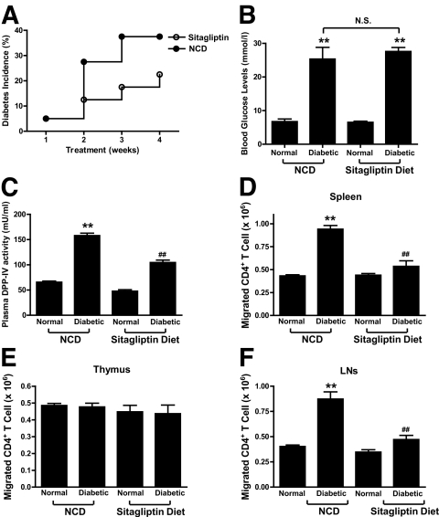
Objective: Treatment of NOD mice with the dipeptidyl peptidase-IV (DPP-IV) inhibitor sitagliptin preserved islet transplants through a pathway involving modulation of splenic CD4(+) T-cell migration. In the current study, effects of sitagliptin on migration of additional subsets of CD4(+) T-cells were examined and underlying molecular mechanisms were further defined.

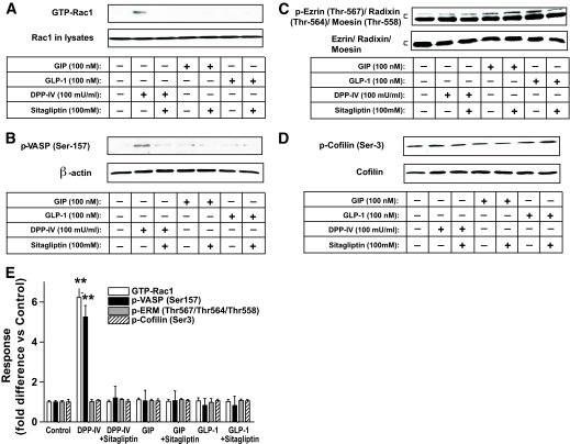
Research design and methods: Effects of sitagliptin on migration of NOD mouse splenic, thymic, and lymph node CD4(+) T-cells were determined. Signaling modules involved in DPP-IV-, Sitagliptin- and incretin-mediated modulation of CD4(+) T-cell migration were studied using Western blot and Rac1 and nuclear factor-kappaB (NF-kappaB) activity assays.

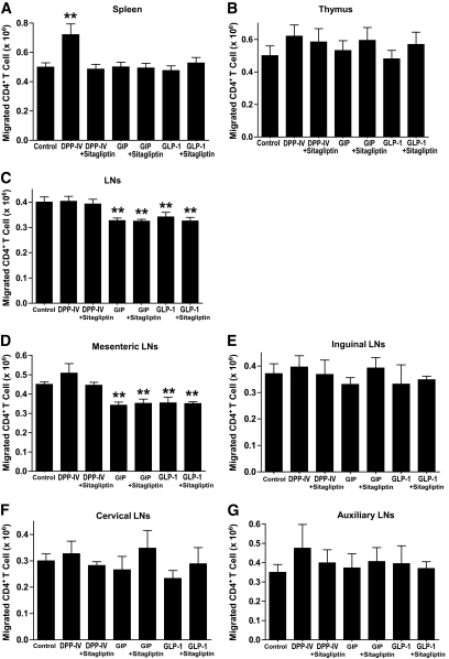
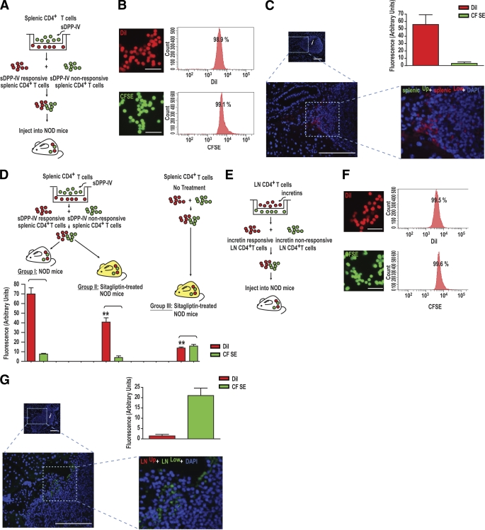
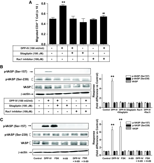
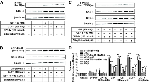
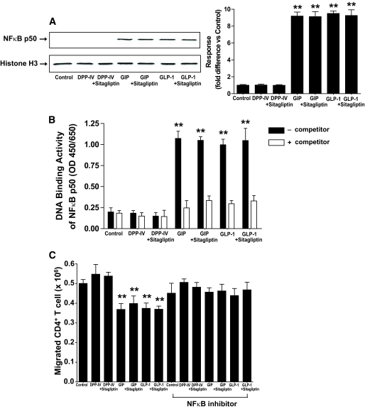
Results: Migration of splenic and lymph node CD4(+) T-cells of diabetic NOD mice was reduced by sitagliptin treatment. In vitro treatment of splenic, but not thymic or lymph node CD4(+) T-cells, from nondiabetic NOD mice with soluble (s) DPP-IV increased migration. Sitagliptin abolished sDPP-IV effects on splenic CD4(+) T-cell migration, whereas incretins decreased migration of lymph node, but not splenic, CD4(+) T-cells. Splenic CD4(+) T-cells demonstrating increased in vitro migration in response to sDPP-IV and lymph node CD4(+) T-cells that were nonresponsive to incretins selectively infiltrated islets of NOD mice, after injection. Sitagliptin decreases migration of splenic CD4(+) T-cells through a pathway involving Rac1/vasodilator-stimulated phosphoprotein, whereas its inhibitory effects on the migration of lymph node CD4(+) T-cells involve incretin-activation of the NF-kappaB pathway.

Conclusions: Benefits of sitagliptin treatment in diabetic NOD mice may be mediated through selective effects on subpopulations of T-cells that are related to autoimmunity.


Figures

FIG. 1.
FIG. 1.
Sitagliptin modulates the migration of splenic and lymph node CD4+ T-cells. Female NOD mice (8–10 weeks old) were fed a NCD or sitagliptin diet for 1 month, before isolation of lymphocytes. A–C: Effects of sitagliptin on incidence of diabetes (A), blood glucose levels (B), and plasma DPP-IV activity (C). d–F: Effect of sitagliptin on the migration of CD4+ T-cells. CD4+ T-cells were isolated from spleen (D), thymus (E), and lymph nodes (F) from the NCD and sitagliptin groups. The migration of CD4+ T-cells was determined using Transwell chamber (Corning) as described in research design and methods. All data are means ± SEM, and significance was tested using ANOVA with a Newman-Keuls post hoc test. **P < 0.05 vs. normal NCD group; ##P < 0.05 vs. diabetic NCD group. N.S., not significant.
FIG. 2.
FIG. 2.
Effects of sDPP-IV and incretins on the migration of CD4+ T-cells. For these and all subsequent studies, CD4+ T-cells were isolated from nondiabetic female NOD mice receiving NCD. Responses of CD4+ T-cells from the spleen (A), thymus (B), and lymph nodes (LNs) (C) stimulated with sDPP-IV (100 mU/ml), GIP, or GLP-1 (100 nmol/l) in the presence or absence of the DPP-IV inhibitor sitagliptin (100 μmol/l) for 1 h. “Control” migration is CD4+ T-cell migration in the absence of sDPP-IV, GIP, or GLP-1. To further delineate the subset of responsive lymph node CD4+ T-cells, cells were isolated from the mesenteric (D), inguinal (E), cervical (F), and auxiliary (G) lymph nodes and treated in an identical fashion to those in (A–C). All data are means ± SEM, and significance was tested using ANOVA with a Newman-Keuls post hoc test. **P < 0.05 vs. control.
FIG. 3.
FIG. 3.
In vivo migration of splenic and lymph node CD4+ T-cells after in vitro treatment with sDPP-IV or incretins. A: Experimental designs for treatment of splenic CD4+ T-cells with sDPP-IV and administration to diabetic NOD mice. Splenic CD4+ T-cells (A) were stimulated for 1 h with sDPP-IV (100 mU/ml). Cells that migrated into the lower compartment were labeled with DiI, and the remaining cells on the upper surface were labeled with CFSE. For studies on incretins, lymph node (LN) CD4+ T-cells (E) were stimulated for 1 h with GIP and GLP-1 (100 nmol/l). Cells that migrated into the lower compartment were labeled with CFSE, and those remaining on the upper surface were labeled with DiI. In both cases, labeled cells were combined and intravenously injected into recipient mice, and pancreata were examined by fluorescent microscopy to detect DiI- or CFSE-labeled CD4+ T-cells in infiltrated islets. B and F: Similar labeling efficiencies of splenic (B) and lymph nodes (F) CD4+ T-cells with DiI or CFSE. Splenic (B) and lymph node (F) CD4+ T-cells were labeled with DiI or CFSE. The labeling was confirmed by fluorescent microscopy, and labeling efficiency was determined by flow cytometry. Shown are representative profiles from n = 3. Scale bar: 10 μm. C and G: Islet localization and quantification of labeled splenic (C) and lymph node (G) CD4+ T-cells after sDPP-IV or incretin stimulation. splenicUp and splenicLow (C) and LNUp and LNLow (G) represent splenic or lymph node lymphocytes from upper and lower chambers, respectively. Mixtures of DiI- and CFSE- labeled splenic (C) and lymph node (G) CD4+ T-cells were intravenously injected into diabetic recipient mice, and pancreatic homing of labeled lymphocytes was determined by confocal fluorescent microscopy. Number of recipient mice: n = 4/group. Infiltrated islets are represented with an i. Scale bar: 50 μm. Upper right: Quantification of recovered fluorescent label from the pancreata. Dye was extracted from pancreata and fluorescence was measured as described in research design and methods. D: Experimental designs for treatment of splenic CD4+ T-cells with sDPP-IV and administration into diabetic NOD mice ± treatment with sitagliptin. The experimental details are as in A, apart from treatment of mice with sitagliptin in groups II and II. Number of recipient mice: n = 6–10/group. Significance was tested using ANOVA with a Newman-Keuls post hoc test. **P < 0.05 vs. group I, DiI. A high-quality digital representation of this figure is available in the online issue.
FIG. 4.
FIG. 4.
Signaling modules potentially involved in the effect of sitagliptin on splenic CD4+ T-cells. Splenic CD4+ T-cells were stimulated in vitro for 1 h with sDPP-IV (100 mU/ml), GIP, or GLP-1 (100 nmol/l) in the presence or absence of DPP-IV inhibitor (100 μmol/l), and total cellular extracts were isolated. A: Rac1 activation. Rac1 activity was determined by Rac1 pulldown assays as described in research design and methods. Briefly, cell lysates were incubated with the agarose-immobilized GST-PAK1, and the coprecipitates were subjected to Western blot hybridization using an anti-Rac1 antibody. Western blot analyses were performed with antibodies against phospho (p)-VASP (Ser157) (B), phospho-Ezrin (Thr567)/Radixin (Thr564)/Moesin (Thr558), Ezrin/Radixin/Moesin (C), phospho-cofilin (Ser3), cofilin (D), and β-actin. E: Densitometric analysis of A–D. All data are means ± SEM, and significance was tested using ANOVA with a Newman-Keuls post hoc test. **P < 0.05 vs. control. Western blots are representative of n = 3.
FIG. 5.
FIG. 5.
Involvement of a Rac1/cAMP/PKA module in sDPP-IV mediated splenic CD4+ T-cell migration and phosphorylation of the cytoskeletal organizing protein VASP. A: Effects of treatment with Rac1 inhibitor on splenic CD4+ T-cell migration. Splenic CD4+ T-cells were stimulated for 1 h with DPP-IV (100 mU/ml) in the presence or absence of Rac1 inhibitor (100 μmol/l), and the migration of CD4+ T-cells was determined. B: Effects of treatment with Rac1 inhibitor on the level of sDPP-IV-mediated phosphorylation of VASP (Ser157) and VASP (Ser239). Splenic CD4+ T-cells were treated as described above, and total cellular extracts were isolated. Western blots were quantified using densitometric analysis and are representative of n = 3. C: Effect of treatment with H-89 on the level of sDPP-IV- or forskolin-mediated phosphorylation of VASP (Ser157) and VASP (Ser239). Splenic CD4+ T-cells were stimulated for 1 h with sDPP-IV (100 mU/ml) or forskolin (FSK; 10 μmol/l) in the presence or absence of H-89 (10 μmol/l). Western blots were quantified using densitometric analysis and are representative of n = 3. All data are means ± SEM, and significance was tested using ANOVA with a Newman-Keuls post hoc test. **P < 0.05 vs. control; ##P < 0.05 vs. DPP-IV.
FIG. 6.
FIG. 6.
Signaling modules potentially involved in the effect of sitagliptin on lymph node CD4+ T-cells. Total cellular extracts were isolated from lymph nodes CD4+ T-cells of nondiabetic female NOD mice placed on NCD and treated as described in the legend to Fig. 3. Western blot analyses were performed with antibodies against phospho (p)-IκB (Ser32), IκB (A), phospho-NF-κB p65 (Ser536), NF-κB p65 (B), phospho-IKKα/β (Ser176/180), IKKα, IKKβ (C), and β-actin. D: Densitometric analysis of A–C. All data represent means ± SEM, and significance was tested using ANOVA with a Newman-Keuls post hoc test. **P < 0.05 vs. control. Western blots are representative of n = 3.
FIG. 7.
FIG. 7.
Involvement of NF-κB activation in the effect of incretins on lymph node CD4+ T-cells. A: Nuclear localization of NF-κB p50. Lymph node CD4+ T-cells were stimulated for 1 h with sDPP-IV (100 mU/ml), GIP, or GLP-1 (100 nmol/l) in the presence or absence of Sitagliptin (100 μmol/l), nuclear extracts were prepared, and Western blot analyses were performed with antibodies against NF-κB p50 and histone H3. Western blots were quantified using densitometric analysis and are representative of n = 3. B: NF-κB p50 transcription factor activation. Nuclear extracts were isolated from lymph node CD4+ T-cells and treated as described above. DNA binding activity of NF-κB p50 transcription factor was determined as described in research design and methods. C: Effect of treatment with NF-κB inhibitor on incretin-mediated lymph node CD4+ T-cell migration. Lymph node CD4+ T-cells were stimulated for 1 h with sDPP-IV (100 mU/ml), GIP, or GLP-1 (100 nmol/l) in the presence or absence of sitagliptin (100 μmol/l) and/or NF-κB inhibitor (7.5 μmol/l). The migration of lymph node CD4+ T-cells was determined using Transwell chambers as described in research design and methods. All data represent means ± SEM, and significance was tested using ANOVA with a Newman-Keuls post hoc test. **P < 0.05 vs. control.

References

    1. Brubaker PL, Drucker DJ: Minireview: Glucagon-like peptides regulate cell proliferation and apoptosis in the pancreas, gut, and central nervous system. Endocrinology 2004;145:2653–2659 - PubMed
    1. Drucker DJ: The role of gut hormones in glucose homeostasis. J Clin Invest 2007;117:24–32 - PMC - PubMed
    1. Yusta B, Baggio LL, Estall JL, Koehler JA, Holland DP, Li H, Pipeleers D, Ling Z, Drucker DJ: GLP-1 receptor activation improves beta cell function and survival following induction of endoplasmic reticulum stress. Cell Metab 2006;4:391–406 - PubMed
    1. Ehses JA, Casilla VR, Doty T, Pospisilik JA, Winter KD, Demuth HU, Pederson RA, McIntosh CH: Glucose-dependent insulinotropic polypeptide promotes beta-(INS-1) cell survival via cyclic adenosine monophosphate-mediated caspase-3 inhibition and regulation of p38 mitogen-activated protein kinase. Endocrinology 2003;144:4433–4445 - PubMed
    1. Kim SJ, Winter K, Nian C, Tsuneoka M, Koda Y, McIntosh CH: Glucose-dependent insulinotropic polypeptide (GIP) stimulation of pancreatic beta-cell survival is dependent upon phosphatidylinositol 3-kinase (PI3K)/protein kinase B (PKB) signaling, inactivation of the forkhead transcription factor Foxo1, and down-regulation of bax expression. J Biol Chem 2005;280:22297–22307 - PubMed

Publication types

MeSH terms