. 2010 Apr 20;107(16):7497-502.
doi: 10.1073/pnas.0907696107. Epub 2010 Apr 5.

IKK{gamma} protein is a target of BAG3 regulatory activity in human tumor growth

Affiliations

IKK{gamma} protein is a target of BAG3 regulatory activity in human tumor growth

Massimo Ammirante et al. Proc Natl Acad Sci U S A. .

Abstract

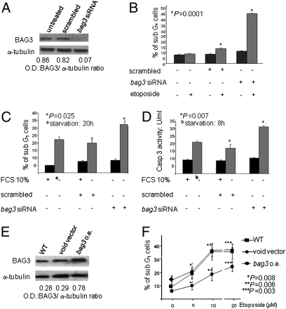
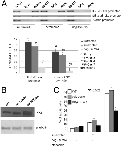
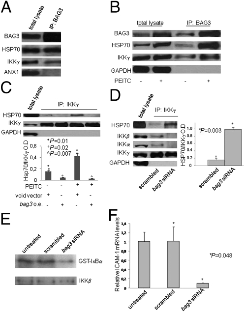
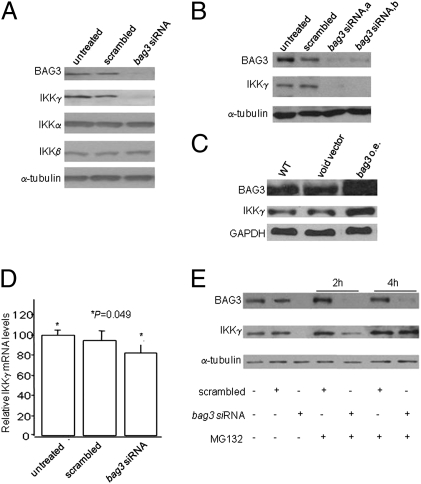
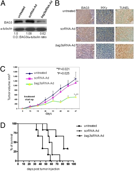
BAG3, a member of the BAG family of heat shock protein (HSP) 70 cochaperones, is expressed in response to stressful stimuli in a number of normal cell types and constitutively in a variety of tumors, including pancreas carcinomas, lymphocytic and myeloblastic leukemias, and thyroid carcinomas. Down-regulation of BAG3 results in cell death, but the underlying molecular mechanisms are still elusive. Here, we investigated the molecular mechanism of BAG3-dependent survival in human osteosarcoma (SAOS-2) and melanoma (M14) cells. We show that bag3 overexpression in tumors promotes survival through the NF-kappaB pathway. Indeed, we demonstrate that BAG3 alters the interaction between HSP70 and IKKgamma, increasing availability of IKKgamma and protecting it from proteasome-dependent degradation; this, in turn, results in increased NF-kappaB activity and survival. These results identify bag3 as a potential target for anticancer therapies in those tumors in which this gene is constitutively expressed. As a proof of principle, we show that treatment of a mouse xenograft tumor model with bag3siRNA-adenovirus that down-regulates bag3 results in reduced tumor growth and increased animal survival.


Conflict of interest statement

The authors declare no conflict of interest.

Figures

Fig. 1.
Fig. 1.
BAG3 protein levels influence starvation or etoposide-induced apoptosis in SAOS-2 cells. (A) SAOS-2 cells (30% confluency) were transfected with bag3siRNA or control scrambled RNA. After 72 h, whole-cell extracts were analyzed by immunoblotting with anti-BAG3 TOS-2 polyclonal antibody or control anti-α-tubulin antibody. (B) Cells were transfected as in A. After 72 h, cells were washed and incubated with medium alone or in the presence of etoposide (20 μM). Twenty-four hours later, apoptosis was evaluated as the percentage of sub-G1 cells by cell permeabilization and propidium iodide staining in flow cytometry. (C) Cells were transfected as in A. After 72 h, cells were washed and incubated with complete medium or in starvation medium (absence of FCS). Twenty hours later, the sub-G1 cell percentage was analyzed. (D) Transfected cells, treated as described in C, were assessed for caspase-3 activity after 8 h of treatment with starvation medium. (E) SAOS-2 cells, WT or transfected (12 weeks with stable transfectants), with a bag3 cDNA construct in either the pcDNA3.1 vector [bag3 overexpressing (o.e.)] or the void vector were analyzed for their content of BAG3 protein by immunoblotting with TOS-2 polyclonal antibody. (F) Cells were transfected as described in E and cultured for 48 h with the indicated concentrations (μM) of etoposide; cell apoptosis was then analyzed by cell permeabilization and PI staining in flow cytometry.
Fig. 2.
Fig. 2.
BAG3 protein levels influence NF-κB activity. (A) SAOS-2 cells at 30% confluency were transfected with bag3 or control scrambled siRNA. Chromatin was immunoprecipitated after 72 h with p65 antibody or control IgGs, and purified DNA was subjected to PCR assay to amplify a segment spanning the κB-responsive elements on the IL-8 and IκB-α promoters. β-actin promoter primers were used as a negative control. The graph depicts band densitometry values from three separate experiments. ns, not significant. (B) Lysates from SAOS-2 cells, WT or transfected (12 weeks of stable transfectants), with either a construct expressing a constitutively active form of IKKβ [IKKβ EE overexpressing (o.e.)] or the void vector were analyzed in immunoblotting with anti-α-tubulin antibody. (C) WT, void vector-transfected, and IKKβ EE-transfected cells were plated at 30% confluency and transfected with scrambled or bag3 siRNA. After 72 h, cells were washed and incubated either with medium alone or in presence of etoposide (20 μM). Twenty-four hours later, apoptosis was analyzed by flow cytometry.
Fig. 3.
Fig. 3.
BAG3 protein levels influence HSP70 association with IKKγ and IKK activity. (A) SAOS-2 cell lysate was immunoprecipitated with an anti-BAG3 polyclonal antibody and analyzed by immunoblotting with anti-HSP70 and anti-IKKγ antibodies. An antibody recognizing annexin I was used as a negative control. (B) M14 whole lysates from untreated or PEITC-treated (5 μM for 30 min) cells were immunoprecipitated with the anti-BAG3 monoclonal antibody AC-2 and analyzed by immunoblotting with anti-BAG3 TOS-2 polyclonal, anti-HSP70, or anti-GAPDH antibody. (C) M14 cells were transfected either with a bag3 overexpressing (o.e.) vector or the void vector. After 48 h, cells were treated as described in B. Immunoprecipitation was performed using an anti-IKKγ polyclonal antibody and analyzed by immunoblotting with anti-HSP70, anti-IKKγ, or anti-GAPDH antibody. The graph depicts densitometry values of bands obtained from two separate experiments. (D) M14 cells were plated at 30% confluency and transfected with scrambled or bag3 siRNA. After 48 h, cell lysates were immunoprecipitated with anti-IKKγ polyclonal antibody and analyzed by immunoblotting with anti-HSP70, anti-IKKγ, anti-IKKα, or anti-IKKβ antibody. The amount of coimmunoprecipitated HSP70 was quantified by densitometry from three separate experiments and normalized to the amount of IKKγ (OD: HSP70/IKKγ). An antibody recognizing GAPDH was used as a negative control. (E) Whole-cell lysates were prepared, and IKK kinase activity was measured after immunoprecipitation with anti-IKKα antibody using GST-IκBα (1–54) as a substrate (34). IKK recovery was determined by immunoblotting with anti-IKKβ antibody. (F) Total RNA was extracted, and ICAM-1 mRNA levels were analyzed by quantitative RT-PCR using β-actin mRNA levels for normalization.
Fig. 4.
Fig. 4.
BAG3 protein influences the intracellular levels of IKKγ protein. (A) SAOS-2 cells (30% confluency) were transfected with scrambled or bag3 siRNA. After 96 h, cell lysates were obtained and analyzed by immunoblotting with anti-BAG3 or anti-α-tubulin antibody. (B) M14 cells were plated at 30% confluency and transfected with two different bag3-specific siRNAs (indicated as bag3 siRNA a and b) or with a control scrambled siRNA. After 96 h, lysates were analyzed by immunoblotting with anti-BAG3 or anti-α-tubulin antibody. (C) M14 cells, WT or transfected, with either a bag3 cDNA construct in pcDNA3.1 vector [bag3 overexpressing (o.e.)] or the void vector were analyzed for their content of BAG3 protein by immunoblotting with TOS-2 polyclonal antibody; furthermore, IKKγ levels were checked by immunoblotting and GAPDH was used to monitor equal loading conditions. (D) Total RNA was extracted, and IKKγ mRNA levels were analyzed by quantitative RT-PCR using 18s rRNA levels for normalization. (E) M14 cells were plated at 30% confluency and transfected with bag3 or control scrambled siRNA. After 96 h, cells were treated with MG132 (10 μM) for 2 and 4 h. Cell lysates were analyzed by immunoblotting with anti-BAG3, anti-IKKγ, or anti-α-tubulin antibody.
Fig. 5.
Fig. 5.
Knockdown of BAG3 protein results in diminishing tumor IKKγ levels, increasing tumor cell apoptosis, inhibiting tumor growth, and enhancing animal survival in M14-xenografted mice. (A) M14 cells (5 × 10⁶) were injected s.c. onto the back of 6-week-old female BALB/c nu/nu mice. Two weeks later, animals were randomized into three groups (10 animals per group) and control PBS (100 μL), bag3siRNA-Ad, or scrRNA-Ad (108 pfu per 100 μL) was injected into the tumors twice a week. After 2 weeks of treatment, four tumors per animal group were excised and BAG3 protein content was analyzed by Western blot. (B) BAG3 and IKKγ protein were detected by immunohistochemistry, whereas apoptosis was analyzed by TUNEL assay, in tumor sections. Representative results are shown. (C) Tumor sizes in bag3siRNA-Ad, scrRNA-Ad, treated, or untreated remaining mice (6 per group) were measured every week using calipers. (D) Evaluation of animal survival was carried out according to Kaplan–Meier analysis. Differences among the treatment groups were analyzed by ANOVA.

References

    1. Thress K, Henzel W, Shillinglaw W, Kornbluth S. Scythe: A novel reaper-binding apoptotic regulator. EMBO J. 1998;17:6135–6143. - PMC - PubMed
    1. Takayama S, Xie Z, Reed JC. An evolutionarily conserved family of Hsp70/Hsc70 molecular chaperone regulators. J Biol Chem. 1999;274:781–786. - PubMed
    1. Briknarová K, et al. Structural analysis of BAG1 cochaperone and its interactions with Hsc70 heat shock protein. Nat Struct Biol. 2001;8:349–352. - PubMed
    1. Moribe Y, Niimi T, Yamashita O, Yaginuma T, Samui A. Samui, a novel cold-inducible gene, encoding a protein with a BAG domain similar to silencer of death domains (SODD/BAG-4), isolated from Bombyx diapause eggs. Eur J Biochem. 2001;268:3432–3442. - PubMed
    1. Takayama S, Reed JC. Molecular chaperone targeting and regulation by BAG family proteins. Nat Cell Biol. 2001;3:237–241. - PubMed

Publication types