. 2010 Apr 20;107(16):7443-8.
doi: 10.1073/pnas.0912943107. Epub 2010 Apr 5.

Embryonic trafficking of gammadelta T cells to skin is dependent on E/P selectin ligands and CCR4

Affiliations

Embryonic trafficking of gammadelta T cells to skin is dependent on E/P selectin ligands and CCR4

Xiaodong Jiang et al. Proc Natl Acad Sci U S A. .

Abstract

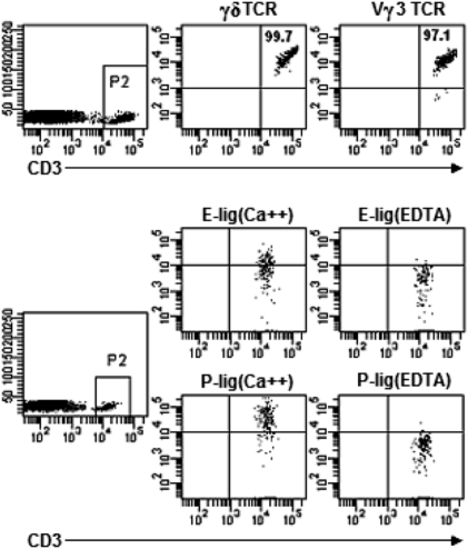
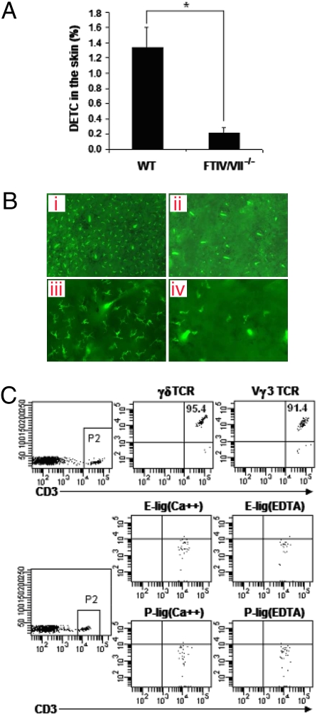
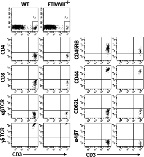
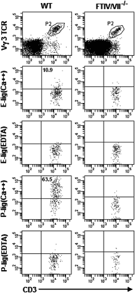
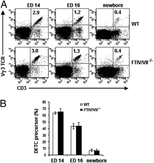
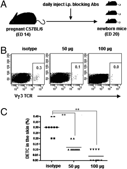
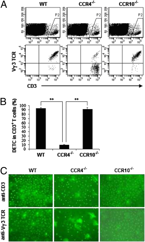
Dendritic epidermal T cells (DETC) express an invariant Vgamma3/Vdelta1 T-cell receptor, appear in fetal epidermis, and form a population of resident epidermal T cells. Their temporal development in the thymus has been studied extensively. However, little is known about the mechanisms involved in the embryonic trafficking of DETC from thymus to epidermis. We demonstrate that DETC in adult skin, as well as the DETC precursors in fetal thymus, express E and P selectin ligands (E- and P-lig). Mice deficient in alpha1,3 fucosyltransferases IV and VII (FTIV/VII) cannot synthesize the carbohydrate motifs that form key elements of these selectin ligands. The numbers of DETC in the epidermis of FTIV/VII(-/-) mice were dramatically reduced compared with normal mice. However, the development of DETC precursors in fetal thymus was identical in normal and FTIV/VII(-/-) mice, suggesting that the DETC precursors produced in FTIV/VII(-/-) mice could not traffic effectively to skin because they lack E- and P-lig. We tested this hypothesis by daily injection of blocking antibodies against E and P selectin into pregnant mice. Mice born from dams treated with anti-selectin antibodies, but not those born from dams treated with isotype control, had significantly diminished numbers of DETC. To test the role of chemokine receptors in DETC skin homing, we examined skin from CCR4(-/-) and CCR10(-/-) mice, respectively. DETC were significantly reduced in CCR4(-/-) mice but were present at normal levels in CCR10(-/-) mice. Our results present evidence for the crucial role of trafficking molecules in embryonic migration of DETC precursors to skin.


Conflict of interest statement

The authors declare no conflict of interest.

Figures

Fig. 1.
Fig. 1.
DETC in normal WT C57BL/6 mice express E-lig and P-lig. Epidermal cell suspensions prepared from 8-week-old WT mice were stained with fluorescence-conjugated anti-CD3, anti-γδ TCR, and anti-Vγ3 TCR, followed with analysis by flow cytometry. The numbers in quadrants indicate the percentages in CD3+ T cells. E- and P-lig expression was examined by incubating skin cells with rmCD62E/Fc or rmCD62P/Fc chimera in HBSS buffer containing 2 mM calcium. HBSS buffer supplemented with 5 mM EDTA was used for the controls. Data are representative of at least three independent experiments.
Fig. 2.
Fig. 2.
DETC are significantly diminished in E-lig/P-lig double-deficient mice. (A) Total skin cells of 8-week-old WT or FTIV/VII−/− C57BL/6 mice were stained with fluorescence-conjugated anti-CD3 and anti-Vγ3 TCR mAbs. The frequency of DETC was analyzed by flow cytometry. *, P < 0.05. (B) Epidermal sheets were peeled from dorsal halves through incubation in 20 mM EDTA for 2–3 h. They then were labeled overnight at 4 °C with FITC-conjugated anti-Vγ3 TCR mAb in PBS containing 2% BSA. Finally, PBS-washed epidermal sheets were mounted on glass slides, and DETC were visualized by immunofluorescence microscope. a and c are WT epidermis sheets; b and d are FTIV/VII−/− epidermis sheets; a and b: low-magnification micrograph (×100); c and d: high-magnification micrograph (×200). (C) E-lig and P-lig expression on DETC of 8-week-old FTIV/VII−/− mice was examined using the binding assay described in Fig. 1. Data are representative of at least three independent experiments.
Fig. 3.
Fig. 3.
Equivalent phenotype of DETC in WT and E-lig/P-lig double-deficient mice. Epidermal cells prepared from 8-week-old WT or FTIV/VII−/− mice were labeled with fluorescence-conjugated mAbs. Given that the vast majority of CD3+ cells in epidermis are Vγ3 TCR+ DETC, the CD3+ cell population thus was gated for DETC phenotype analysis.
Fig. 4.
Fig. 4.
Thymic DETC precursors in normal WT C57BL/6 mice express E-lig and P-lig. Fetal thymocyte suspensions of WT or FTIV/VII−/− mice at ED15 were prepared. E-lig and P-lig expression was examined using the binding assay described in Fig. 1. The numbers in quadrants indicate the percentages in DETC precursors.
Fig. 5.
Fig. 5.
E-lig/P-lig double deficiency does not impair DETC development in the fetal thymus. (A) Fetal or newborn thymocyte suspensions prepared at various EDs (above plots) were labeled with fluorescence-conjugated anti-CD3 and anti-Vγ3 TCR mAbs. Results are representative of three experiments at each time-point. The numbers in quadrants indicate the percentages of DETC precursors of total thymocytes. (B) Summary data (mean ± SD) on the frequency of DETC precursors of CD3+ thymocytes. Profiles are representative of three independent experiments.
Fig. 6.
Fig. 6.
Blocking E selectin and P selectin in vivo reduces DETC number in neonatal skin. (A) Experimental design. From ED14 to ED20 (birth), the pregnant WT C57BL/6 mice were injected i.p. daily with combined mAbs (100 μg or 50 μg each) against E selectin and P selectin or with isotype-matched control mAbs. The neonatal back skins were harvested for the preparation of single-cell suspensions by enzymatic digestion. (B) Cells then were labeled for DETC frequency analysis by flow cytometry. (C) Summary data on the percentages of DETC in neonatal skin after blocking E selectin and P selectin. **, P < 0.01. Results are representative of two independent experiments.
Fig. 7.
Fig. 7.
DETC are diminished in CCR4−/− but not in CCR10−/− mice. (A) Epidermal cells were prepared from 8-week-old WT, CCR4−/−, or CCR10−/− C57BL/6 mice. Cells then were washed thoroughly and labeled with fluorescence-conjugated anti-CD3 and anti-Vγ3 TCR mAbs for DETC analysis by flow cytometry. (B) Summary data on the percentages of DETC in CD3+ T cells. **, P < 0.01. (C) DETC on epidermal sheets were visualized by labeling with FITC-conjugated anti-CD3 or anti-Vγ3 TCR mAb (×100). One of three independent experiments with similar results is shown.

References

    1. Saito H, et al. Complete primary structure of a heterodimeric T-cell receptor deduced from cDNA sequences. Nature. 1984;309:757–762. - PubMed
    1. Hayday AC, et al. Structure, organization, and somatic rearrangement of T cell gamma genes. Cell. 1985;40:259–269. - PubMed
    1. Brenner MB, et al. Identification of a putative second T-cell receptor. Nature. 1986;322:145–149. - PubMed
    1. Nanno M, Shiohara T, Yamamoto H, Kawakami K, Ishikawa H. gammadelta T cells: Firefighters or fire boosters in the front lines of inflammatory responses. Immunol Rev. 2007;215:103–113. - PubMed
    1. Payer E, Elbe A, Stingl G. Epidermal T lymphocytes—ontogeny, features and function. Springer Semin Immunopathol. 1992;13:315–331. - PubMed

Publication types

LinkOut - more resources