. 2010 Apr 20;107(16):7449-54.
doi: 10.1073/pnas.0909707107. Epub 2010 Apr 5.

Metallofullerene nanoparticles circumvent tumor resistance to cisplatin by reactivating endocytosis

Affiliations

Metallofullerene nanoparticles circumvent tumor resistance to cisplatin by reactivating endocytosis

Xing-Jie Liang et al. Proc Natl Acad Sci U S A. .

Abstract

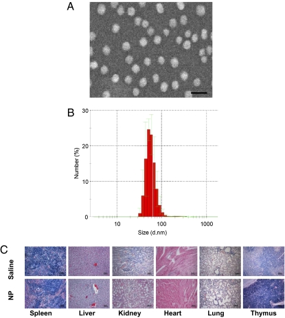
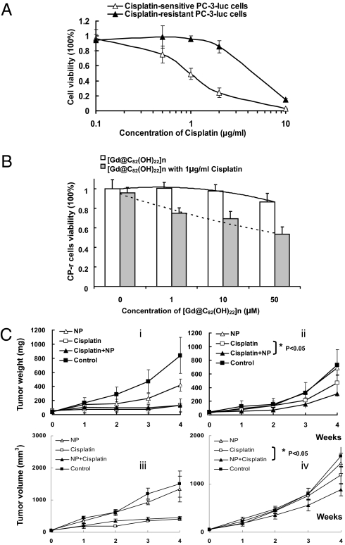
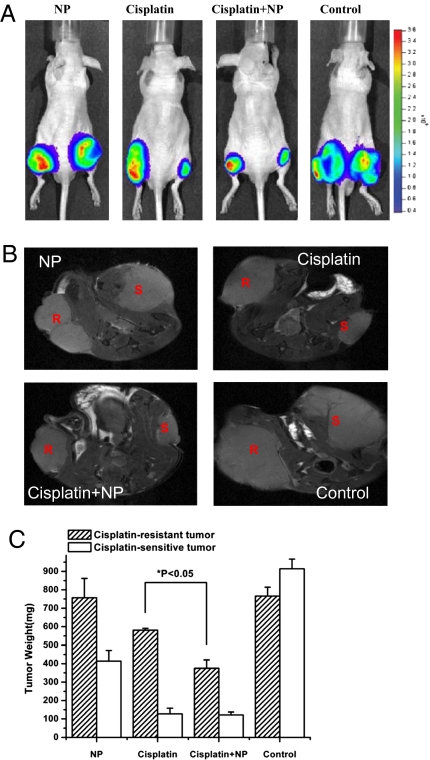
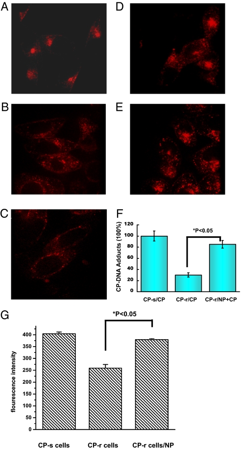
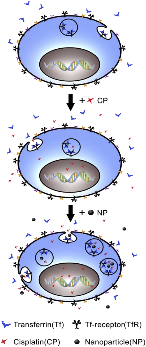
Cisplatin is a chemotherapeutic drug commonly used in clinics. However, acquired resistance confines its application in chemotherapeutics. To overcome the acquired resistance to cisplatin, it is reasoned, based on our previous findings of mediation of cellular responses by [Gd@C(82)(OH)(22)](n) nanoparticles, that [Gd@C(82)(OH)(22)](n) may reverse tumor resistance to cisplatin by reactivating the impaired endocytosis of cisplatin-resistant human prostate cancer (CP-r) cells. Here we report that exposure of the CP-r PC-3-luc cells to cisplatin in the presence of nontoxic [Gd@C(82)(OH)(22)](n) not only decreased the number of surviving CP-r cells but also inhibited growth of the CP-r tumors in athymic nude mice as measured by both optical and MRI. Labeling the CP-r PC-3 cells with transferrin, an endocytotic marker, demonstrated that pretreatment of the CP-r PC-3-luc cells with [Gd@C(82)(OH)(22)](n) enhanced intracellular accumulation of cisplatin and formation of cisplatin-DNA adducts by restoring the defective endocytosis of the CP-r cancer cells. The results suggest that [Gd@C(82)(OH)(22)](n) nanoparticles overcome tumor resistance to cisplatin by increasing its intracellular accumulation through the mechanism of restoring defective endocytosis. The technology can be extended to other challenges related to multidrug resistance often found in cancer treatments.


Conflict of interest statement

The authors declare no conflict of interest.

Figures

Fig. 1.
Fig. 1.
Characterization of [Gd@C82(OH)22]n nanoparticles. (A) [Gd@C82(OH)22]n nanoparticles characterized by scanning electron microscopy (SEM). (Scale bar, 100 nm.) (B) Size of [Gd@C82(OH)22]n nanoparticles, as measured by dynamic light scattering (DSL). (C) Representative histological H&E staining of various organ tissues from mice treated with [Gd@C82(OH)22]n nanoparticles or saline. (Scale bar, 50 μm.)
Fig. 2.
Fig. 2.
[Gd@C82(OH)22]n nanoparticles induced sensitivity enhancement of CP-r cells and tumors to cisplatin. (A) Cellular viability curves of CP-s and CP-r PC-3-luc cells treated with cisplatin. Cells (3 × 10³) were plated in 96-well plates with 100 μL medium per well. After 6 h, various concentrations of cisplatin in 50 μL medium were added, and the cells were incubated at 37 °C for 3 days. Before the cell viability measurement, 10 μL of MTT solution (Kumamoto) was added to each well and incubated for 2 h. Cell viability was measured using spectrophotometry. (B) Measurement of [Gd@C82(OH)22]n nanoparticle cytotoxicity in CP-r cells treated or untreated with cisplatin (1 μg/mL). The protocol was the same as described in Fig. 2A. Results shown are the average of three different experiments. (C) Tumor weight was measured by caliper quantification {tumor weight (mg) = tumor density (1 mg/mm3) × length (mm) × [width (mm)2 / 2]} (i, CP-s PC-3-luc; ii, CP-r PC-3-luc). Nanoparticles (20 μM) were used. Tumor volumes were derived from consecutive multiple MRI images using Image J software (iii, CP-s PC-3-luc; iv, CP-r PC-3-luc). Statistical analysis of Inset C using ANOVA shows a significant difference between two sets of data when P < 0.05.
Fig. 3.
Fig. 3.
Sensitizing CP-r tumors to cisplatin treatment by [Gd@C82(OH)22]n nanoparticles in vivo. (A) Optical imaging for comparison of the sizes of CP-s and CP-r PC-3-luc tumors treated with either [Gd@C82(OH)22]n nanoparticles (NP, 20 μM), cisplatin, cisplatin plus nanoparticles (cisplatin + NP), or saline solution alone as a control. (B) MRI images of CP-s and CP-r PC-3-luc tumors after 4 weeks of various treatments described in A. (Right) CP-s tumor. (Left) CP-r tumor. (C) Weights of tumors were measured at the end of 4 weeks’ treatment. MRI images were analyzed by Image J software for tumor volume. Tumor weight was calculated by conversion of volume to weight. NP (20 μM) treatment significantly enhanced the ability of cisplatin to inhibit growth of CP-r tumors.
Fig. 4.
Fig. 4.
[Gd@C82(OH)22]n nanoparticles increased transferrin-mediated endo-cytosis. Texas Red–transferrin conjugates were used to label CP-s and CP-r PC-3-luc cells for endocytotic measurement. Cisplatin concentration was 1 μg/mL, and [Gd@C82(OH)22]n was 20 μM. (A–E) Confocal microscopy images. (A) CP-s cells. (B) CP-r cells. (C) CP-r cells treated with cisplatin. (D) CP-r cells treated with nanoparticles. (E) CP-r cells treated with cisplatin and nanoparticles. (F) ICP-MS/MS measurements of DNA adducts in CP-s treated cisplatin; CP-r cells treated with cisplatin with/without [Gd@C82(OH)22]n. Data presented are the integrated ICP-MS signals as mean values of yield measurements for various DNA adducts measured by ICP-MS in cells after various treatments. (G) CP-s and CP-r cells were labeled with Texas Red–transferrin for endocytotic measurement. Fluorescence intensity of internalized Texas Red–transferrin was measured by spectrophotometer at an excitation wavelength of 595 nm and emission wavelength of 620 nm. Results shown are mean of three different experiments. Comparisons between groups were evaluated by one-way ANOVA. There was a statistically significant difference between the CP-R cells and CP-R cells treated with nanoparticles.
Fig. 5.
Fig. 5.
[Gd@C82(OH)22]n nanoparticles enhance CP-r cell sensitivity to cisplatin. (Top) Normal endocytosis that includes binding of ligands (e.g., transferrin: Tf) to their Tf-receptors on plasma membrane followed by binding, ingestion into cytoplasma, intracellular vesicle transportation, payload release, and vesicle recycle. (Middle) Receptor-mediated endocytosis of cisplatin in the CP-r cells. Because of defective endocytosis, there is less intracellular accumulation of cisplatin and therefore less formation of cisplatin-DNA adducts in the CP-r cells. (Bottom) Nanoparticle-activated endocytosis in the CP-r cells, resulting in more efficient transportation of cisplatin-containing vesicles and more cisplatin binding to nucleic acid to sensitize the CP-r cells.

References

    1. Gottlieb JA, Drewinko B. Review of the current clinical status of platinum coordination complexes in cancer chemotherapy. Cancer Chemother Rep. 1975;59:621–628. - PubMed
    1. Wang D, Lippard SJ. Cellular processing of platinum anticancer drugs. Nat Rev Drug Discov. 2005;4:307–320. - PubMed
    1. Borst P, Rottenberg S, Jonkers J. How do real tumors become resistant to cisplatin? Cell Cycle. 2008;7:1353–1359. - PubMed
    1. Siddik ZH. Cisplatin: Mode of cytotoxic action and molecular basis of resistance. Oncogene. 2003;22:7265–7279. - PubMed
    1. Kartner N, Riordan JR, Ling V. Cell surface P-glycoprotein associated with multidrug resistance in mammalian cell lines. Science. 1983;221:1285–1288. - PubMed

Publication types

LinkOut - more resources