. 2010 Apr 20;121(15):1735-45.
doi: 10.1161/CIRCULATIONAHA.109.899252. Epub 2010 Apr 5.

Human adult vena saphena contains perivascular progenitor cells endowed with clonogenic and proangiogenic potential

Affiliations

Human adult vena saphena contains perivascular progenitor cells endowed with clonogenic and proangiogenic potential

Paola Campagnolo et al. Circulation. .

Abstract

Background: Clinical trials in ischemic patients showed the safety and benefit of autologous bone marrow progenitor cell transplantation. Non-bone marrow progenitor cells with proangiogenic capacities have been described, yet they remain clinically unexploited owing to their scarcity, difficulty of access, and low ex vivo expansibility. We investigated the presence, antigenic profile, expansion capacity, and proangiogenic potential of progenitor cells from the saphenous vein of patients undergoing coronary artery bypass surgery.

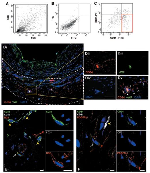
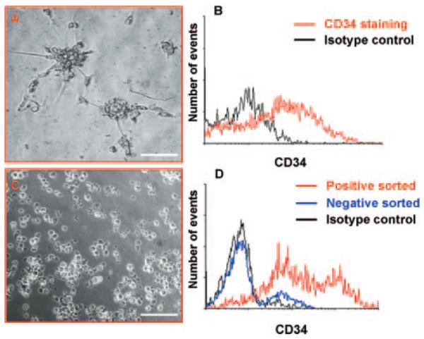
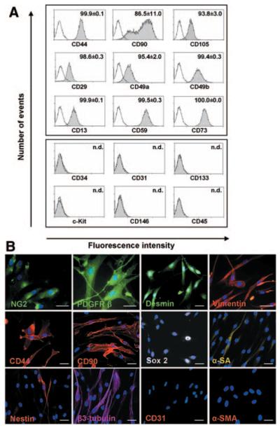
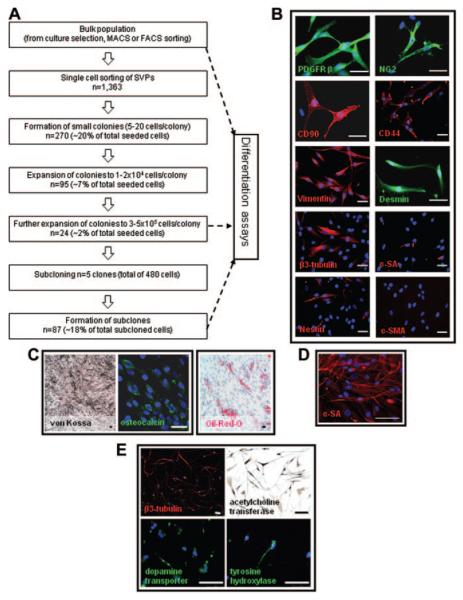
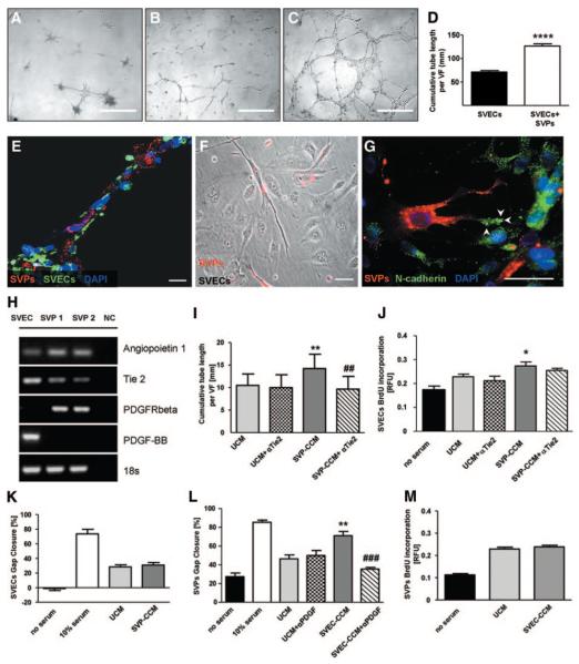
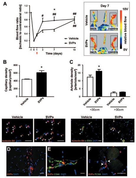
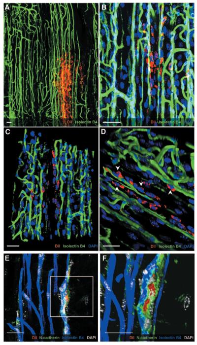
Methods and results: CD34-positive cells, negative for the endothelial marker von Willebrand factor, were localized around adventitial vasa vasorum. After dissection of the vein from surrounding tissues and enzymatic digestion, CD34-positive/CD31-negative cells were isolated by selective culture, immunomagnetic beads, or fluorescence-assisted cell sorting. In the presence of serum, CD34-positive/CD31-negative cells gave rise to a highly proliferative population that expressed pericyte/mesenchymal antigens together with the stem cell marker Sox2 and showed clonogenic and multilineage differentiation capacities. We called this population "saphenous vein-derived progenitor cells" (SVPs). In culture, SVPs integrated into networks formed by endothelial cells and supported angiogenesis through paracrine mechanisms. Reciprocally, endothelial cell-released factors facilitated SVP migration. These interactive responses were inhibited by Tie-2 or platelet-derived growth factor-BB blockade. Intramuscular injection of SVPs in ischemic limbs of immunodeficient mice improved neovascularization and blood flow recovery. At 14 days after transplantation, proliferating SVPs were still detectable in the recipient muscles, where they established N-cadherin-mediated physical contact with the capillary endothelium.

Conclusions: SVPs generated from human vein CD34-positive/CD31-negative progenitor cells might represent a new therapeutic tool for angiogenic therapy in ischemic patients.


Figures

Figure 1
Figure 1
Identification of CD34pos cells in saphenous veins. Representative scattergrams (A and B) showing the relative abundance of CD34posCD31neg cells (C, red box) within saphenous vein digests. Forward-sideward scatterplot (A) and isotype control are shown (B). FSC indicates forward scatter; SSC, side scatter; PE, phycoerythrin; and FITC, fluorescein isothiocyanate. Di, Low-magnification picture shows morphology of saphenous vein and distribution of vasa vasorum in adventitia (delimited by dashed lines). Sections were stained for progenitor/EC marker CD34 (red; Dii), EC marker vWF (green; Diii), and nuclei (DAPI, blue, Div). Representative confocal image merge reveals the presence of CD34posvWFneg progenitor cells located in the perivascular zone of the vasa vasorum (white arrowheads in Dv). Furthermore, in the same region, a few CD34pos cells (green; E and F) coexpressed NG2 (red, E) or PDGFRβ (red, F). Double-positive cells are indicated by white arrows. CD31 (white; E and F) stained ECs of vasa vasorum (yellow arrowheads; E and F). Higher-magnification panels in E and F show adventitial CD34pos cells, negative for CD31 and coexpressing NG2 or PDGFRβ. Scale bar=50 μm (D) and 10 μm (E and F).
Figure 2
Figure 2
Isolation of CD34pos cells. Morphology of spheroids derived from saphenous vein cells cultured for 5 days in HM (A). Spheroids were stained with CD34 or isotype control. Flow cytometry showed that they comprised ≈50% of CD34pos cells (B). Cells derived from magnetic bead isolation appeared as floating single cells (C). Histogram shows flow cytometry analysis of cells enriched by isolation with magnetic beads (D). Representatives of n=5 (A and B) and n=6 (C and D) experiments. Scale bar=100 μm.
Figure 3
Figure 3
Characterization of the CD34posCD31neg-derived population. After culture in serum-containing medium, CD34posCD31neg cells differentiated into cells expressing mesenchymal/pericyte markers as assessed by flow cytometry (A; n.d. indicates not detectable; white, isotype control, gray, specific antibody staining [as indicated]) and immunocytochemistry (B; scale bar=50 μm, nuclei: DAPI [blue]). α-SA indicates α-sarcomeric actin; α-SMA, α-smooth muscle actin. Results are representative of 4 (A) or 3 (B) experiments.
Figure 4
Figure 4
Clonogenic assay and differentiation of SVPs. Single SVPs (1363) were deposited on Terasaki plates. Of the seeded cells, ≈20% formed small colonies, and 2% gave rise to large colonies and could be subcloned (efficiency 18%; A). Clones conserved the mesenchymal/pericyte phenotype of the polyclonal population (B). Under appropriate differentiation conditions, SVPs, their clones, and subclones displayed a wide differentiation capacity, which gave rise to cells of mesodermal lineage such as osteoblasts, adipocytes (C), and myocytes (D; α-sarcomeric actin, [α-SA]). They also generated neuron-like cells (E). Nuclei are identified by DAPI (blue). Scale bar 50 μm (B and C) or 100 μm (D and E). α-SMA indicates α-smooth muscle actin.
Figure 5
Figure 5
Interactions between SVPs and SVECs. SVPs formed peculiar structures in Matrigel (A). Network formation was strongly enhanced in SVP-SVEC coculture (C) compared with culture of SVECs alone (B). D, Bar graph showing average tube length; values are mean±SEM of 4 experiments performed in quadruplicate. VF indicates view field. ****P<0.0001 vs SVECs. Confocal microscopy image shows tubes formed by SVECs (isolectin, green) and covered by SVPs (DiI, red; E). F, Phase-contrast microscopy image showing the interaction of SVPs (DiI, red) with multiple SVECs (unstained) in 2D coculture. G, Representative confocal microscopy image showing different patterns of N-cadherin staining (green fluorescence) in SVPs (localized at filopodia, white arrowheads) and ECs (diffuse to all of the cell membrane). Scale bars: 500 μm (A, B, and C); 50 μm (F and G); 20 μm (E). H, Expression of Ang-1/Tie-2 and PDGF-BB/PDGFRβ in SVECs and SVPs. NC indicates negative control. I through M, Bar graphs show the reciprocal paracrine effect of SVECs and SVP-CCM. SVP-CCM influenced SVEC network-formation capacity (I) and proliferation (J) but not migration (K). Tie-2 blockade inhibited the stimulatory action on the network-formation capacity of SVECs (I). SVP-CCM enhanced SVEC migration (L), this effect being inhibited by PDGF-BB blockade, but did not alter their proliferation (M). Positive control was medium that contained 10% serum, whereas negative control was serum-deprived medium. Values are mean±SEM of 3 experiments performed in quadruplicate. *P<0.05 and **P<0.01 vs unconditioned culture medium; ##P<0.01 and ###P<0.001 vs CCM. UCM indicates unconditioned medium; BrdU, bromodeoxyuridine; and RFU, relative fluorescence unit.
Figure 6
Figure 6
In vivo proangiogenic effect of SVPs. A, Line graphs and representative laser Doppler images taken at day 7 from induction of unilateral limb ischemia (isch) illustrating the improvement of blood flow recovery in SVP-treated compared with vehicle-injected mice (arrow indicates time of cell injection; n=7 animals per group). Contra indicates contralateral. B and C, Bar graphs and representative pictures show increased capillary (B) and arteriole (C) density in muscles injected with SVPs compared with vehicle at 14 days after ischemia. Values are mean±SEM; n=4 animals per group.*P<0.05 vs vehicle; #P<0.05 and ##P<0.01 vs day 1. SVPs (DiI, red) persisted in ischemic muscle 14 days after injection (D), expressed human CD44 (E), and were positive for proliferating cell nuclear antigen (F; PCNA, arrowheads). Scale bar=20 μm. α-SMA indicates α-smooth muscle actin.
Figure 7
Figure 7
Interaction of SVPs with the host vasculature. Z-stack of confocal images showing numerous SVPs (CM-DiI; red) surrounding the host vasculature (isolectin B4; green; A and B). 3D reconstruction of the field shows physical contact between SVPs and ECs (C and D; white arrowheads). Direct interaction was also confirmed by N-cadherin staining (green), which accumulated in the junction between SVPs (red) and capillary ECs (blue; E and F). Nuclei were stained with DAPI (blue in A through D; white in E and F). Scale bar=25 μm.

References

    1. Howson KM, Aplin AC, Gelati M, Alessandri G, Parati EA, Nicosia RF. The postnatal rat aorta contains pericyte progenitor cells that form spheroidal colonies in suspension culture. Am J Physiol Cell Physiol. 2005;289:C1396–C1407. - PubMed
    1. Invernici G, Emanueli C, Madeddu P, Cristini S, Gadau S, Benetti A, Ciusani E, Stassi G, Siragusa M, Nicosia R, Peschle C, Fascio U, Colombo A, Rizzuti T, Parati E, Alessandri G. Human fetal aorta contains vascular progenitor cells capable of inducing vasculogenesis, angiogenesis, and myogenesis in vitro and in a murine model of peripheral ischemia. Am J Pathol. 2007;170:1879–1892. - PMC - PubMed
    1. Zengin E, Chalajour F, Gehling UM, Ito WD, Treede H, Lauke H, Weil J, Reichenspurner H, Kilic N, Ergun S. Vascular wall resident progenitor cells: a source for postnatal vasculogenesis. Development. 2006;133:1543–1551. - PubMed
    1. Barcelos LS, Duplaa C, Krankel N, Graiani G, Invernici G, Katare R, Siragusa M, Meloni M, Campesi I, Monica M, Simm A, Campagnolo P, Mangialardi G, Stevanato L, Alessandri G, Emanueli C, Madeddu P. Human CD133+ progenitor cells promote the healing of diabetic ischemic ulcers by paracrine stimulation of angiogenesis and activation of Wnt signaling. Circ Res. 2009;104:1095–1102. - PMC - PubMed
    1. Beltrami AP, Cesselli D, Bergamin N, Marcon P, Rigo S, Puppato E, D'Aurizio F, Verardo R, Piazza S, Pignatelli A, Poz A, Baccarani U, Damiani D, Fanin R, Mariuzzi L, Finato N, Masolini P, Burelli S, Belluzzi O, Schneider C, Beltrami CA. Multipotent cells can be generated in vitro from several adult human organs (heart, liver, and bone marrow) Blood. 2007;110:3438–3446. - PubMed

Publication types

MeSH terms

Substances