. 2010 Apr 12;207(4):699-709.
doi: 10.1084/jem.20091618. Epub 2010 Apr 5.

OX40 is required for regulatory T cell-mediated control of colitis

Affiliations

OX40 is required for regulatory T cell-mediated control of colitis

Thibault Griseri et al. J Exp Med. .

Abstract

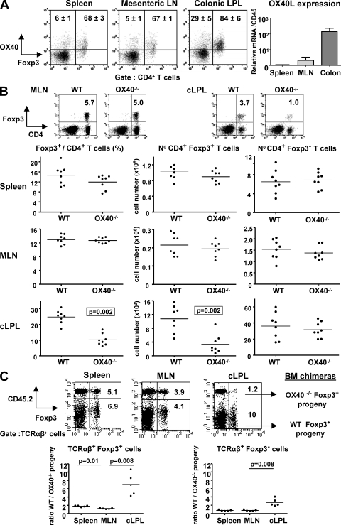
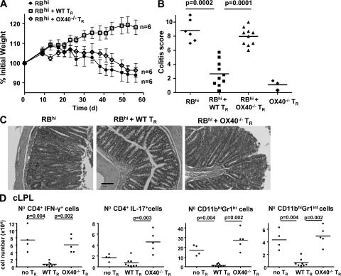
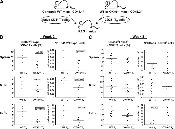
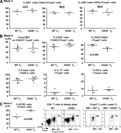
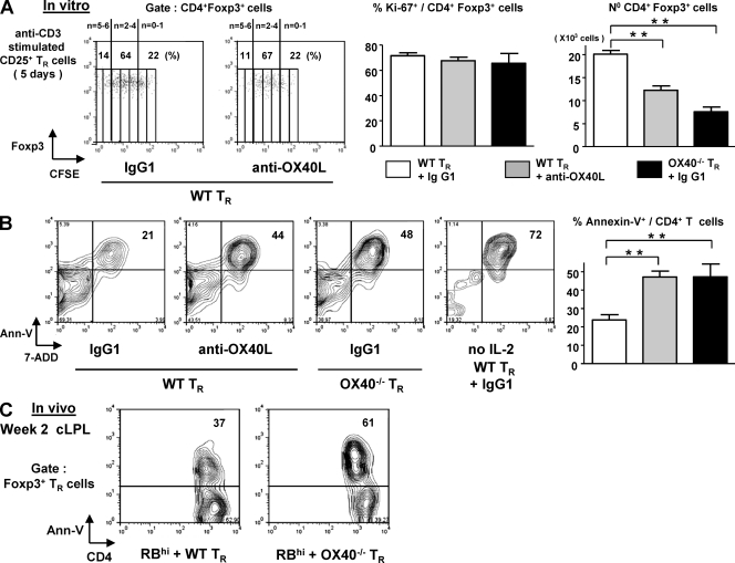
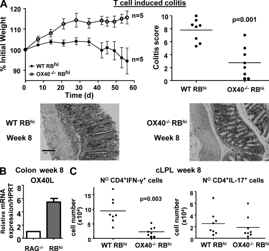
The immune response in the gastrointestinal tract is a tightly controlled balance between effector and regulatory cell responses. Here, we have investigated the role of OX40 in influencing the balance between conventional T cells and Foxp3+ regulatory T (T reg) cells. Under steady-state conditions, OX40 was required by T reg cells for their accumulation in the colon, but not peripheral lymphoid organs. Strikingly, under inflammatory conditions OX40 played an essential role in T reg cell-mediated suppression of colitis. OX40(-/-) T reg cells showed reduced accumulation in the colon and peripheral lymphoid organs, resulting in their inability to keep pace with the effector response. In the absence of OX40 signaling, T reg cells underwent enhanced activation-induced cell death, indicating that OX40 delivers an important survival signal to T reg cells after activation. As OX40 also promoted the colitogenic Th1 response, its expression on T reg cells may be required for effective competition with OX40-dependent effector responses. These results newly identify a key role for OX40 in the homeostasis of intestinal Foxp3+ T reg cells and in suppression of colitis. These fi ndings should be taken into account when considering OX40 blockade for treatment of IBD.


Figures

Figure 1.
Figure 1.
OX40 is preferentially expressed on intestinal T cells and promotes Foxp3+ T reg cell accumulation in the colon in a cell intrinsic way. (A, left) Tissues from WT mice analyzed by flow cytometry for CD4, Foxp3, and OX40 expression. Percentages of OX40+ cells among CD4+ Foxp3 or Foxp3+ cells are indicated in representative dot plots (n = 5). (right) OX40L mRNA amounts in spleen, MLN, and total colon homogenate (n = 5). Values are normalized to CD45 and are mean values (± SEM) relative to spleen (set to 1). (B) Percentages and total numbers of Foxp3 and Foxp3+ CD4 T cells in tissues of WT and OX40−/− mice. (C) BM cells from WT congenic mice (CD45.1+) mixed in equal proportion with BM cells from OX40−/− mice (CD45.2+) were injected i.v. into irradiated RAG−/− mice. 8 wk after transfer, tissues were harvested and stained for TCR-β, CD45.2, and Foxp3. Representative staining (top) and the ratio of WT/OX40−/− progeny among TCRαβ+ cells are shown (bottom). Each point represents an individual mouse and horizontal bars represent group means (B and C). Data are representative of two to three independents experiments.
Figure 2.
Figure 2.
OX40 expression on T reg cells is required to suppress T cell induced colitis. RAG−/− mice received WT CD4+CD45RBhi T cells alone (no TR), or in combination with WT or OX40−/− CD4+CD25+ T reg cells (TR). (A) Mean weight of six animals per group (± SEM). (B) Colitis score at 8 wk. An additional control group showed that OX40−/− T reg cells do not induce colitis by themselves. (C) Photomicrographs of mid-colon sections. Bar, 100 µm. (D) Number of colonic IFN-γ+ and IL-17+CD4+ T cells, neutrophils (CD11cCD11bhiGr1hiSSChi) and monocytes/macrophages (CD11cCD11bhiGr1intSSClo) at 8 wk. Each point represents an individual mouse and horizontal bars represent group means (B and D). Results are representative of three independent experiments.
Figure 3.
Figure 3.
OX40 is required for early Foxp3+ T reg cell accumulation in the lymphoid organs and colon during control of intestinal inflammation. (A) CD45.1+ CD4+CD45RBhi T cells were cotransferred with CD45.2+ WT or OX40−/− T reg cells (TR) into RAG−/− mice. Numbers and percentages of CD45.2+CD4+Foxp3+ cells in tissues 2 wk (B) and 8 wk (C) after cotransfer. Each point represents an individual mouse. Data are representative of three independent experiments.
Figure 4.
Figure 4.
Phenotypic and functional characteristics of WT and OX40−/− T reg cells after transfer. CD45.1+ CD4+CD45RBhi T cells were transferred with CD45.2+ WT or OX40−/− T reg cells (TR) into RAG−/− mice. (A) Ki67 expression in cLPL and MLN and α4β7 integrin expression in MLN on Foxp3+ T reg progeny at wk 2. (B) Foxp3, CD25, CD44, CD62L expression, IL-17, and IFN-γ secretion by Foxp3+ T reg progeny at wk 8 in cLPL. (C) GITR expression by Foxp3+ T reg progeny at 2 wk (dot plots) and 8 wk (graph) and CD4+ T cells from the spleen of normal mice (dot plots). Percentages of GITRhi cells among CD4+Foxp3+ cells and GITR MFI on Foxp3+GITR+cells are indicated in dot plots. Data are representative of two independent experiments.
Figure 5.
Figure 5.
Lack of an OX40 signal renders activated T reg cells more susceptible to apoptosis. WT or OX40−/− CD25+ FACS sorted T reg cells were cultured in triplicate for 5 d with irradiated APC and soluble anti-CD3, in the presence of an anti-OX40L mAb or isotype control. hIL-2 was added to the cultures (200U/ml). (A) Representative dot plots of CFSE dilution by CD4+Foxp3+ cells. Bar graphs represent means of Ki67 expression by Foxp3+ cells and total number of CD4+Foxp3+ cells at d 5 (mean of triplicate cultures ± SD). (B) Representative Annexin V-7 ADD staining 5 d after T reg cell activation (gated on CD4+ T cells). Bar graph represents mean percentages of apoptotic cells (mean of triplicate cultures ± SD). **, P < 0.001. Results are representatives of two to three independent experiments. (C) CD4+CD45RBhi T cells were transferred into RAG−/− mice with CD25+ TR cells isolated from WT or OX40−/− Foxp3-GFP mice. Numbers in dot plots represent the percentages of Annexin V+ cells at 2 wk among colonic CD4+Foxp3(GFP)+ cells, after overnight culture in medium. Three to four colons were pooled per group, and results are representative of two independent experiments.
Figure 6.
Figure 6.
OX40 is required for the development of colitogenic T cell responses. RAG−/− mice received CD4+CD45RBhi T cells from WT or OX40−/− donors. (A) Mean weight loss (n = 6 ± SEM) and colitis score at wk 8. Representative photomicrographs of mid-colon sections are shown below. Bar, 100 µm. (B) Amount of OX40L mRNA in total colon homogenate from untransferred mice (RAG−/−) or colitic mice (RBhi) at wk 8. Data shown are the mean values (n = 4) relative to untransferred RAG−/− mice (set to 1), after normalization to HPRT expression. (C) Number of colonic IFN-γ+ and IL-17+ CD4+ T cells 8 wk after transfer. Each point in (A, right) and (C) represents an individual mouse. Results are representative of three independent experiments.

References

    1. Bennett C.L., Christie J., Ramsdell F., Brunkow M.E., Ferguson P.J., Whitesell L., Kelly T.E., Saulsbury F.T., Chance P.F., Ochs H.D. 2001. The immune dysregulation, polyendocrinopathy, enteropathy, X-linked syndrome (IPEX) is caused by mutations of FOXP3. Nat. Genet. 27:20–21 10.1038/83713 - DOI - PubMed
    1. Bouma G., Strober W. 2003. The immunological and genetic basis of inflammatory bowel disease. Nat. Rev. Immunol. 3:521–533 10.1038/nri1132 - DOI - PubMed
    1. Cederbom L., Hall H., Ivars F. 2000. CD4+CD25+ regulatory T cells down-regulate co-stimulatory molecules on antigen-presenting cells. Eur. J. Immunol. 30:1538–1543 10.1002/1521-4141(200006)30:6<1538::AID-IMMU1538>3.0.CO;2-X - DOI - PubMed
    1. Chaudhry A., Rudra D., Treuting P., Samstein R.M., Liang Y., Kas A., Rudensky A.Y. 2009. CD4+ Regulatory T cells control TH17 responses in a Stat3-dependent manner. Science. 326:986–991 - PMC - PubMed
    1. Chen Q., Davidson T.S., Huter E.N., Shevach E.M. 2009. Engagement of TLR2 does not reverse the suppressor function of mouse regulatory T cells, but promotes their survival. J. Immunol. 183:4458–4466 10.4049/jimmunol.0901465 - DOI - PMC - PubMed

Publication types

MeSH terms