

The role of osteopontin and tumor necrosis factor alpha receptor-1 in xenobiotic-induced cholangitis and biliary fibrosis in mice
- PMID: 20368698
- PMCID: PMC4285781
- DOI: 10.1038/labinvest.2010.61
The role of osteopontin and tumor necrosis factor alpha receptor-1 in xenobiotic-induced cholangitis and biliary fibrosis in mice
Abstract
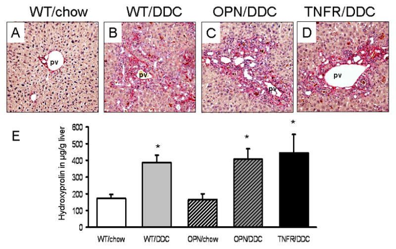
Proinflammatory and profibrotic cytokines such as osteopontin (OPN) and tumor necrosis factor-alpha receptor-1 (TNFR(1)) may be critically involved in the pathogenesis of cholangiopathies and biliary fibrosis. We therefore aimed to determine the role of genetic loss of either OPN or TNFR(1) in 3,5-diethoxycarbonyl-1,4-dihydrocollidine (DDC)-fed mice as a model of xenobiotic-induced sclerosing cholangitis with biliary-type liver fibrosis using respective knock-out mice. OPN and TNFR(1) knock-out mice were fed a 0.1% DDC-supplemented diet for 4 weeks and compared with corresponding wild-type (WT) controls. Liver morphology (H&E staining), serum markers of liver injury and cholestasis (ALT, AP, bilirubin), markers of inflammation in liver (CD11b and F4/80 immunostaining, mRNA expression of iNOS, MCP-1, IL-1beta, INF-gamma, TNF-alpha and OPN), degree of ductular reaction (immunohistochemistry with morphometric analysis and western blotting for cholangiocyte-specific marker keratin 19) and degree of liver fibrosis (Sirius-red staining, hepatic hydroxyproline content for quantification) were compared between groups. DDC feeding in OPN and TNFR(1) knock-out mice and respective WT controls resulted in comparable extent of liver injury, inflammatory response, ductular reaction and liver fibrosis. Our data indicate that genetic loss of neither OPN nor TNFR(1) significantly effects on the pathogenesis of DDC-induced sclerosing cholangitis, ductular reaction and resulting biliary fibrosis.
Figures


Similar articles
-
A new xenobiotic-induced mouse model of sclerosing cholangitis and biliary fibrosis.Am J Pathol. 2007 Aug;171(2):525-36. doi: 10.2353/ajpath.2007.061133. Epub 2007 Jun 28. Am J Pathol. 2007. PMID: 17600122 Free PMC article.
-
Effects of Melittin Treatment in Cholangitis and Biliary Fibrosis in a Model of Xenobiotic-Induced Cholestasis in Mice.Toxins (Basel). 2015 Aug 25;7(9):3372-87. doi: 10.3390/toxins7093372. Toxins (Basel). 2015. PMID: 26308055 Free PMC article.
-
Nuclear Translocation of RELB Is Increased in Diseased Human Liver and Promotes Ductular Reaction and Biliary Fibrosis in Mice.Gastroenterology. 2019 Mar;156(4):1190-1205.e14. doi: 10.1053/j.gastro.2018.11.018. Epub 2018 Nov 13. Gastroenterology. 2019. PMID: 30445013
-
Xenobiotic-induced liver injury and fibrosis.Expert Opin Drug Metab Toxicol. 2012 May;8(5):571-80. doi: 10.1517/17425255.2012.674511. Epub 2012 Mar 28. Expert Opin Drug Metab Toxicol. 2012. PMID: 22452290 Review.
-
Animal models of cholestasis: An update on inflammatory cholangiopathies.Biochim Biophys Acta Mol Basis Dis. 2019 May 1;1865(5):954-964. doi: 10.1016/j.bbadis.2018.07.025. Epub 2018 Aug 11. Biochim Biophys Acta Mol Basis Dis. 2019. PMID: 30398152 Review.
Cited by
-
Animal models of biliary injury and altered bile acid metabolism.Biochim Biophys Acta Mol Basis Dis. 2018 Apr;1864(4 Pt B):1254-1261. doi: 10.1016/j.bbadis.2017.06.027. Epub 2017 Jul 11. Biochim Biophys Acta Mol Basis Dis. 2018. PMID: 28709963 Free PMC article. Review.
-
Osteopontin: an indispensable component in common liver, pancreatic, and biliary related disease.J Cancer Res Clin Oncol. 2024 Nov 22;150(12):508. doi: 10.1007/s00432-024-06038-0. J Cancer Res Clin Oncol. 2024. PMID: 39572438 Free PMC article. Review.
-
Mammalian Target of Rapamycin Complex 2 Signaling Is Required for Liver Regeneration in a Cholestatic Liver Injury Murine Model.Am J Pathol. 2020 Jul;190(7):1414-1426. doi: 10.1016/j.ajpath.2020.03.010. Epub 2020 Apr 7. Am J Pathol. 2020. PMID: 32275903 Free PMC article.
-
A preliminary in vivo study of the effects of OPN on rat liver regeneration induced by partial hepatectomy.Mol Biol Rep. 2016 Dec;43(12):1371-1382. doi: 10.1007/s11033-016-4071-2. Epub 2016 Sep 1. Mol Biol Rep. 2016. PMID: 27585571
-
FXR controls CHOP expression in steatohepatitis.FEBS Lett. 2017 Oct;591(20):3360-3368. doi: 10.1002/1873-3468.12845. Epub 2017 Oct 11. FEBS Lett. 2017. PMID: 28895119 Free PMC article.
References
-
- Lazaridis KN, Strazzabosco M, LaRusso NF. The cholangiopathies: disorders of biliary epithelia. Gastroenterology. 2004;127(5):1565–1577. - PubMed
-
- Lu BR, Mack CL. Inflammation and biliary tract injury. Curr Opin Gastroenterol. 2009;25(3):260–4. - PubMed
-
- Strazzabosco M, Fabris L, Spirli C. Pathophysiology of cholangiopathies. J Clin Gastroenterol. 2005;39(4 Suppl 2):S90–S102. - PubMed
-
- Xia X, Demorrow S, Francis H, et al. Cholangiocyte injury and ductopenic syndromes. Semin Liver Dis. 2007;27(4):401–12. - PubMed
-
- Zatloukal K, Stumptner C, Fuchsbichler A, et al. The keratin cytoskeleton in liver diseases. J Pathol. 2004;204(4):367–76. - PubMed
Publication types
MeSH terms
Substances
Grants and funding
LinkOut - more resources
Full Text Sources
Other Literature Sources
Medical
Molecular Biology Databases
Research Materials
Miscellaneous
