

S100A1: a regulator of striated muscle sarcoplasmic reticulum Ca2+ handling, sarcomeric, and mitochondrial function
- PMID: 20368797
- PMCID: PMC2846685
- DOI: 10.1155/2010/178614
S100A1: a regulator of striated muscle sarcoplasmic reticulum Ca2+ handling, sarcomeric, and mitochondrial function
Abstract
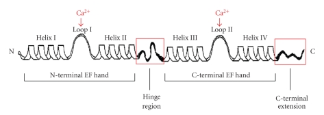
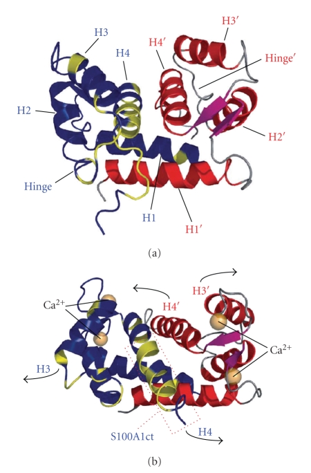
Calcium (Ca(2+)) signaling plays a key role in a wide range of physiological functions including control of cardiac and skeletal muscle performance. To assure a precise coordination of both temporally and spatially transduction of intracellular Ca(2+) oscillations to downstream signaling networks and target operations, Ca(2+) cycling regulation in muscle tissue is conducted by a plethora of diverse molecules. Ca(2+) S100A1 is a member of the Ca(2+)-binding S100 protein family and represents the most abundant S100 isoform in cardiac and skeletal muscle. Early studies revealed distinct expression patterns of S100A1 in healthy and diseased cardiac tissue from animal models and humans. Further elaborate investigations uncovered S100A1 protein as a basic requirement for striated muscle Ca(2+) handling integrity. S100A1 is a critical regulator of cardiomyocyte Ca(2+) cycling and contractile performance. S100A1-mediated inotropy unfolds independent and on top of beta AR-stimulated contractility with unchanged beta AR downstream signaling. S100A1 has further been detected at different sites within the cardiac sarcomere indicating potential roles in myofilament function. More recently, a study reported a mitochondrial location of S100A1 in cardiomyocytes. Additionally, normalizing the level of S100A1 protein by means of viral cardiac gene transfer in animal heart failure models resulted in a disrupted progression towards cardiac failure and enhanced survival. This brief review is confined to the physiological and pathophysiological relevance of S100A1 in cardiac and skeletal muscle Ca(2+) handling with a particular focus on its potential as a molecular target for future therapeutic interventions.
Figures

Similar articles
-
S100A1 in cardiovascular health and disease: closing the gap between basic science and clinical therapy.J Mol Cell Cardiol. 2009 Oct;47(4):445-55. doi: 10.1016/j.yjmcc.2009.06.003. Epub 2009 Jun 16. J Mol Cell Cardiol. 2009. PMID: 19538970 Free PMC article. Review.
-
S100A1: a regulator of myocardial contractility.Proc Natl Acad Sci U S A. 2001 Nov 20;98(24):13889-94. doi: 10.1073/pnas.241393598. Proc Natl Acad Sci U S A. 2001. PMID: 11717446 Free PMC article.
-
S100A1: a novel inotropic regulator of cardiac performance. Transition from molecular physiology to pathophysiological relevance.Am J Physiol Regul Integr Comp Physiol. 2007 Aug;293(2):R568-77. doi: 10.1152/ajpregu.00075.2007. Epub 2007 Apr 25. Am J Physiol Regul Integr Comp Physiol. 2007. PMID: 17459908 Review.
-
S100A1: a multifaceted therapeutic target in cardiovascular disease.J Cardiovasc Transl Res. 2010 Oct;3(5):525-37. doi: 10.1007/s12265-010-9211-9. Epub 2010 Jul 20. J Cardiovasc Transl Res. 2010. PMID: 20645037 Free PMC article. Review.
-
Cardiac AAV9-S100A1 gene therapy rescues post-ischemic heart failure in a preclinical large animal model.Sci Transl Med. 2011 Jul 20;3(92):92ra64. doi: 10.1126/scitranslmed.3002097. Sci Transl Med. 2011. PMID: 21775667 Free PMC article.
Cited by
-
Review of RyR1 pathway and associated pathomechanisms.Acta Neuropathol Commun. 2016 Nov 17;4(1):121. doi: 10.1186/s40478-016-0392-6. Acta Neuropathol Commun. 2016. PMID: 27855725 Free PMC article. Review.
-
Molecular Basis of S100A1 Activation and Target Regulation Within Physiological Cytosolic Ca2+ Levels.Front Mol Biosci. 2020 Jun 23;7:77. doi: 10.3389/fmolb.2020.00077. eCollection 2020. Front Mol Biosci. 2020. PMID: 32656226 Free PMC article.
-
Increased serum levels of S100A1, ZAG, and adiponectin in cachectic patients with COPD.Int J Chron Obstruct Pulmon Dis. 2018 Oct 8;13:3157-3163. doi: 10.2147/COPD.S172996. eCollection 2018. Int J Chron Obstruct Pulmon Dis. 2018. PMID: 30349224 Free PMC article.
-
Calprotectin (S100A8/S100A9): a key protein between inflammation and cancer.Inflamm Res. 2018 Oct;67(10):801-812. doi: 10.1007/s00011-018-1173-4. Epub 2018 Aug 6. Inflamm Res. 2018. PMID: 30083975 Review.
-
S100 proteins in cardiovascular diseases.Mol Med. 2023 May 22;29(1):68. doi: 10.1186/s10020-023-00662-1. Mol Med. 2023. PMID: 37217870 Free PMC article. Review.
References
-
- Bers DM. Cardiac excitation-contraction coupling. Nature. 2002;415(6868):198–205. - PubMed
-
- Most P, Remppis A, Pleger ST, Katus HA, Koch WJ. S100A1: a novel inotropic regulator of cardiac performance. Transition from molecular physiology to pathophysiological relevance. American Journal of Physiology. 2007;293(2):R568–R577. - PubMed
-
- Marenholz I, Heizmann CW, Fritz G. S100 proteins in mouse and man: from evolution to function and pathology (including an update of the nomenclature) Biochemical and Biophysical Research Communications. 2004;322(4):1111–1122. - PubMed
-
- Donato R. Intracellular and extracellular roles of S100 proteins. Microscopy Research and Technique. 2003;60(6):540–551. - PubMed
Publication types
MeSH terms
Substances
Grants and funding
LinkOut - more resources
Full Text Sources
Research Materials
Miscellaneous
