. 2010 Jan 30;3(1):55-68.

Tissue-microarray based immunohistochemical analysis of survival pathways in nodular sclerosing classical Hodgkin lymphoma as compared with Non-Hodgkin's lymphoma

Affiliations

Tissue-microarray based immunohistochemical analysis of survival pathways in nodular sclerosing classical Hodgkin lymphoma as compared with Non-Hodgkin's lymphoma

Jitakshi De et al. Int J Clin Exp Med. .

Abstract

Neoplastic cells rely on key oncogenic pathways for their gain of proliferative and/or loss of apoptotic potential. Therapy targeted at specific points in these pathways has the potential to eliminate cancer cells by inducing differentiation or apoptosis. Concurrent immunophenotypic evaluation of survival pathways in nodular sclerosing classical Hodgkin lymphoma (cHL-NS) tissues has not previously been undertaken. We took the tissue microarray (TMA)-based approach to retrospectively evaluate the activation state of key oncogenic pathways by immunohisto-chemistry (IHC) in a series of 6 cases of cHL-NS (with predominantly syncitial areas). For comparison, 2 cases of diffuse large B-cell lymphoma (DLBCL), and 1 case of follicular hyperplasia (FH) were included in the study. Infiltration of T regulatory cells (Tregs) in the tumor microenvironment was assessed by expression of the Foxp3 transcription factor. Differential upregulation of the mitogen-activated protein kinase (MAPK)-extracellular signal related kinase (ERK), signal transducers and activators of transcription (STAT)3, and protein kinase c - alpha (PKC-alpha) pathways was seen among the cHL cases, whereas nuclear factor - kappa B (NF-kB) and phosphoinositide 3 kinase (PI3 K)-AKT-mammalian target of rapamycin (mTOR) pathways were equally activated in the neoplastic Reed-Sternberg cells of the 6 cHL-NS cases. Marked difference in the morphoproteomic profile was seen amongst the two cases of DLBCL. The amount of Foxp3+ T regulatory cells (Tregs) in the tumor microenvironment was highly variable ranging from 6/hpf to 120/hpf in cHL-NS, and 1/hpf to 82/hpf in DLBCL. In this pilot study, concurrent evaluation of oncogenic pathways in cHL-NS and DLBCL offers powerful insights in the putative therapeutic targets for an individualized approach to diagnosis and therapy.

Keywords: Classical Hodgkin lymphoma; diffuse large B-cell lymphoma; morphoproteomics; survival pathways; tissue microarray.


Figures

Figure 1
Figure 1
Survival pathways with their cross-talk and immune regulatory mechanisms are shown in this pictorial representation of the Reed-Sternberg (R-S) cell. CD30, CD40, and receptor activator of NF-kB (RANK) receptors on R-S cells are activated by autocrine and paracrine loops leading to activation of PI3K and phosphorylation of AKT, promoting growth and survival. IL-21 activates STAT3 and upregulates STAT3 target genes, triggering anti-apoptotic mechanisms, and inducing trafficking of CCR4 and CCR6- expressing T regulatory cells towards the neoplastic cells. Phos-phorylated STAT3 upregulates IL-23 and induces Foxp3 expression in Tregs expressing the IL-23 receptor. These immune regulatory cells modulate the anti-tumor immunity by secretion of IL-10 and IFN-y, both of which block the adaptive and effector immune response. PKC-α activation is associated with increased immune modulation and reduced apoptosis by Bcl-2 overexpression in hematologic malignancies. Inhibition of AKT, NF-kB, STAT3, PKC-α and MAPK-ERK pathways induce cell cycle arrest and apoptosis. The use of an SFK inhibitor has the potential to modulate the immune escape mechanisms by blocking the STAT pathway.
Figure 2
Figure 2
Outlay of the tissue microarray constructed with 9 (2mm) cylindrical cores from representative paraffin-embedded tissue blocks. H&E at original magnification.
Figure 3
Figure 3
Histologic representation of the 9 cases included in this study. 1-6) nodular sclerosing classical Hodgkin lymphoma with predominantly syncitial areas, 7) diffuse large B-cell lymphoma with CD5-positivity, 8) diffuse large B-cell lymphoma with Bcl-6 positivity and concomitant Grade 1-2 follicular lymphoma, 9) reactive lymph node with follicular hyperplasia. H&E × 40X.
Figure 4
Figure 4
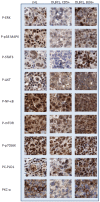
Tissue expression profile of a representative case of classical Hodgkin lymphoma is shown here. The choice of markers was based on current literature of constitutively activate pathways in classical Hodgkin lymphoma (NF-kB, PI3-AKT-mTOR, MAPK-ERK, STAT-3) and PKC-α which promotes growth in hematologic malignancies. Nuclear p-STAT3, p-NF-kB, p-ERK, p-p38 MAPK is evidence of activation of these pathways in majority of cHL cases evaluated in this study. Phosphorylated ERK is differentially expressed in the neoplastic cells of classical Hodgkin lymphoma. Intense membranous PKC-α was noted in the majority of cHL cases with PLD1 co-localization. Original magnification x 400X.
Figure 5
Figure 5
Infiltration of T regulatory cells within the tumor microenvironment is shown here by immunostaining for Foxp3 transcription factor. Enumeration of the number of Foxp3+ cells demonstrates variability amongst the classical Hodgkin lymphoma cases (see Table 2), and distinct difference amongst the two cases of DLBCL with the GC-derived lymphoma (#8) surrounded by greater number of T regulatory cells than the ABC-derived lymphoma (#7) and the reactive lymph node (#9). Original magnification x 200X.
Figure 6
Figure 6
Tissue expression profile of proteins within key survival pathways is shown with comparison amongst a representative cHL case and the two cases of DLBCL. While p-NF-kB, p-mTOR, and p-p70 S6K were nuclear in all three examples, nuclear p-AKT was lacking in the two cases of DLBCL as opposed to cHL which showed nuclear localization of p-AKT (suggesting mTORC1 complex is operative in DLBCL, and mT0RC2 complex in cHL). PKC-α was intensely reactive in cHL and Bcl6+ DLBCL, but not in CD5+ DLBCL. The Bcl-6+ DLBCL (#8) differed from the CD5+ DLBCL (#7) included in the study in its higher p-p38 MAPK, p-ERK, p-STAT3 and PKC-α reactivity; and lower p-NF-kB expression. Original magnification x 200X.

References

    1. Diehl V, Thomas RK, Re D. Part II: Hodgkin's lymphoma-diagnosis and treatment. Lancet Oncol. 2004;5:19–26. - PubMed
    1. Dancey J, Sausville EA. Issues and progress with protein kinase inhibitors for cancer treatment. Nat Rev Drug Discov. 2003;2:296–313. - PubMed
    1. Hinz M, Loser P, Mathas S, Krappmann D, Dorken B, Scheidereit C. Constitutive NF-kappaB maintains high expression of a characteristic gene network, including CD40, CD86, and a set of antiapoptotic genes in Hodgkin/Reed-Sternberg cells. Blood. 2001;97:2798–2807. - PubMed
    1. Brown RE, Kamal NR. p-p70S6K (Thr 389) Expression in nodular sclerosing hodgkin's disease as evidence for receptor tyrosine kinase signaling. Ann Clin Lab Sci. 2005;35:413–414. - PubMed
    1. Kube D, Holtick U, Vockerodt M, Ahmadi T, Haier B, Behrmann I, Heinrich PC, Diehl V, Tesch H. STAT3 is constitutively activated in Hodgkin cell lines. Blood. 2001;98:762–770. - PubMed

LinkOut - more resources