

Effect of phosphate treatment of Acid-etched implants on mineral apposition rates near implants in a dog model
- PMID: 20369085
- PMCID: PMC2946350
Effect of phosphate treatment of Acid-etched implants on mineral apposition rates near implants in a dog model
Abstract
Purpose: This study evaluated the effects of phosphate coating of acid-etched titanium on the mineral apposition rate (MAR) and new bone-to-implant contact (BIC) in a canine model.
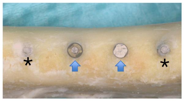
Materials and methods: Titanium implants (2.2 3 4 mm) with acid-etched surfaces that were electrolytically phosphated or not were placed in 48 mandibular sites in six foxhounds. Tetracycline and calcein dyes were administered 1 week after implant placement and 1 week before sacrifice. At 12 weeks after implant placement, the animals were sacrificed. MAR and BIC were evaluated using fluorescence microscopy. Light microscopic and histologic evaluations were performed on undecalcified sections.
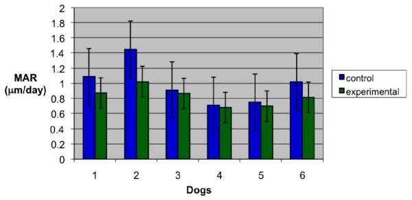
Results: Microscopic evaluation showed the presence of healthy osteoblasts lining bone surfaces near implants. Similar BIC was observed in phosphated and nonphosphated titanium implant sites. MAR was significantly higher around the nonphosphated titanium implant surfaces than around the phosphated titanium samples. No significant differences were found between dogs or implant sites.
Conclusion: Acid-etched implants showed significantly higher MARs compared to acid-etched, phosphate-coated implants. Int J Maxillofac Implants 2010;25:278-286.
Figures


Similar articles
-
Effect of dehiscences to the bone response of implants with an Acid-etched surface: an experimental study in miniature pigs.J Oral Implantol. 2011;37(1):3-17. doi: 10.1563/AAID-JOI-D-09-00090. Epub 2010 Jun 17. J Oral Implantol. 2011. PMID: 20557147
-
Bone mineral apposition rates at early implantation times around differently prepared titanium surfaces: a study in beagle dogs.Int J Oral Maxillofac Implants. 2011 Jan-Feb;26(1):63-9. Int J Oral Maxillofac Implants. 2011. PMID: 21365039
-
Surface-conditioned dental implants: an animal study on bone formation.J Clin Periodontol. 2009 Oct;36(10):882-91. doi: 10.1111/j.1600-051X.2009.01466.x. Epub 2009 Sep 7. J Clin Periodontol. 2009. PMID: 19735467
-
A pilot histologic comparison of bone-to-implant contact between phosphate-coated and control titanium implants in the canine model.Int J Oral Maxillofac Implants. 2014 Jan-Feb;29(1):203-10. doi: 10.11607/jomi.3364. Int J Oral Maxillofac Implants. 2014. PMID: 24451872
-
Comparison of the effects of phosphate-coated and sandblasted acid-etched titanium implants on osseointegration: a microcomputed tomographic examination in the canine model.Int J Oral Maxillofac Implants. 2012 Sep-Oct;27(5):1069-80. Int J Oral Maxillofac Implants. 2012. PMID: 23057019
Cited by
-
Osseointegration behavior of novel Ti-Nb-Zr-Ta-Si alloy for dental implants: an in vivo study.J Mater Sci Mater Med. 2016 Sep;27(9):139. doi: 10.1007/s10856-016-5755-9. Epub 2016 Aug 17. J Mater Sci Mater Med. 2016. PMID: 27534399
-
Reliability of new poly (lactic-co-glycolic acid) membranes treated with oxygen plasma plus silicon dioxide layers for pre-prosthetic guided bone regeneration processes.Med Oral Patol Oral Cir Bucal. 2017 Mar 1;22(2):e242-e250. doi: 10.4317/medoral.21512. Med Oral Patol Oral Cir Bucal. 2017. PMID: 28160588 Free PMC article.
-
A systematic review on the effect of inorganic surface coatings in large animal models and meta-analysis on tricalcium phosphate and hydroxyapatite on periimplant bone formation.J Biomed Mater Res B Appl Biomater. 2022 Jan;110(1):157-175. doi: 10.1002/jbm.b.34899. Epub 2021 Jul 16. J Biomed Mater Res B Appl Biomater. 2022. PMID: 34272804 Free PMC article.
References
-
- Seckinger RJ, Barber HD, Phillips K, Saleh N, Ferarie J. A clinical study of titanium plasma sprayed (TPS)-coated threaded and TPS-coated cylindrical endosseous dental implants. Guide Impl Res. 1996;1:5–8.
-
- Bränemark PI. Vital microscopy of bone marrow in rabbit. Diss Lund Scand J Lab Invest Suppl. 1959;3811:1–82. - PubMed
-
- Schroeder A, van der Zypen E, Stich H, Sutter F. The reactions of bone, connective tissue, and epithelium to endosteal implants with titanium-sprayed surfaces. J Maxillofac Surg. 1981;9:15–25. - PubMed
-
- Bränemark PI, Zarb GA, Albrektsson T. Introduction to Osseointegration. In: Bränemark PI, Zarb GA, Albrektsson T, editors. Tissue Integrated Prostheses: Osseointegration in Clinical Dentistry. Quintessence; Chicago: 1995.
-
- Albrektsson T. In: Tissue Integrated Prostheses: Osseointegration in Clinical Dentistry. Bränemark PI, Zarb GA, Albrektsson T, editors. Quintessence; Chicago: 1985.
Publication types
MeSH terms
Substances
Grants and funding
LinkOut - more resources
Full Text Sources
Miscellaneous