. 2010 Aug;67(16):2787-98.
doi: 10.1007/s00018-010-0364-0. Epub 2010 Apr 6.

Ctenidins: antimicrobial glycine-rich peptides from the hemocytes of the spider Cupiennius salei

Affiliations

Ctenidins: antimicrobial glycine-rich peptides from the hemocytes of the spider Cupiennius salei

Tommy Baumann et al. Cell Mol Life Sci. 2010 Aug.

Abstract

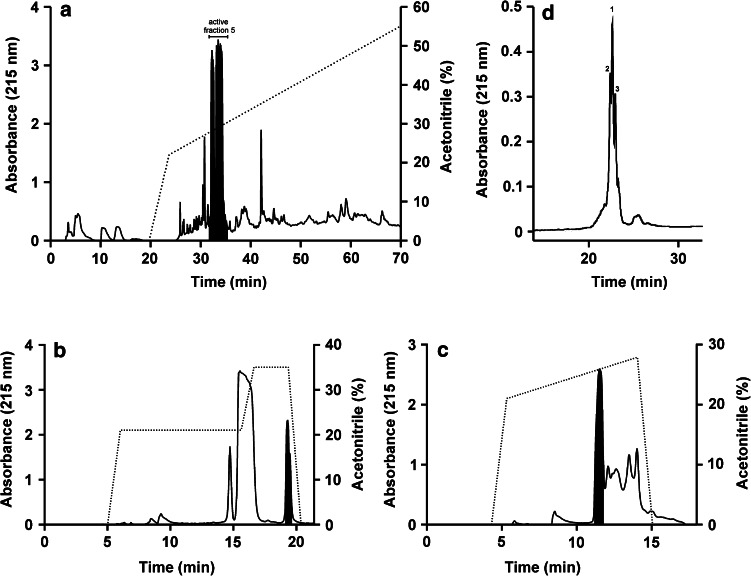
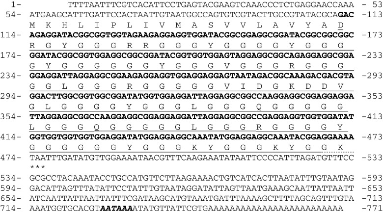
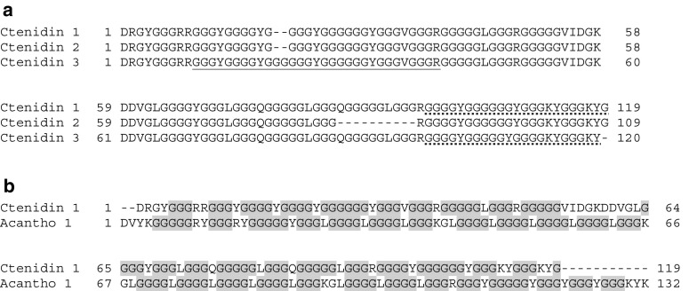
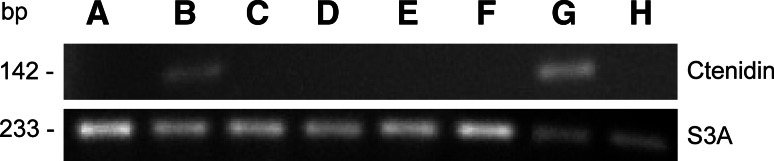
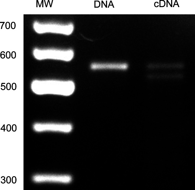
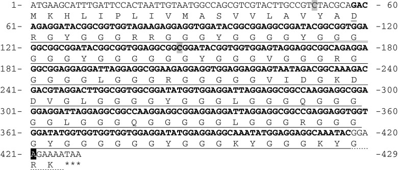
Three novel glycine-rich peptides, named ctenidin 1-3, with activity against the Gram-negative bacterium E. coli, were isolated and characterized from hemocytes of the spider Cupiennius salei. Ctenidins have a high glycine content (>70%), similarly to other glycine-rich peptides, the acanthoscurrins, from another spider, Acanthoscurria gomesiana. A combination of mass spectrometry, Edman degradation, and cDNA cloning revealed the presence of three isoforms of ctenidin, at least two of them originating from simple, intronless genes. The full-length sequences of the ctenidins consist of a 19 amino acid residues signal peptide followed by the mature peptides of 109, 119, or 120 amino acid residues. The mature peptides are post-translationally modified by the cleavage of one or two C-terminal cationic amino acid residue(s) and amidation of the newly created mature C-terminus. Tissue expression analysis revealed that ctenidins are constitutively expressed in hemocytes and to a small extent also in the subesophageal nerve mass.


Figures

Fig. 1
Fig. 1
Purification of ctenidins from C. salei hemocytes. a Preparative RP-HPLC chromatogram of hemocyte lysates. The area in black corresponds to fraction number 5, which showed activity against E. coli and S. aureus. b, c Further separation of the bactericidal active fraction (black areas) was done by two subsequent preparative RP-HPLC purification steps. d Small scale RP-HPLC chromatogram of the active purified sub-fraction containing the ctenidins. The numbers of the peaks correspond to the numbering of the different ctenidins
Fig. 2
Fig. 2
Ctenidin 1 and 2 cDNA sequences. The deduced amino acid sequence is presented under the nucleotide sequence. The mature peptide sequence is underlined; the part missing in ctenidin 2 is double underlined. The nucleotide sequence of the mature peptide is in bold. The dotted line marks the post-translationally cleaved off lysine and the glycine residue involved in C-terminal amidation of the other glycine. Asterisks mark the stop codon. The polyadenylation signal is shown in bold italics
Fig. 3
Fig. 3
Comparisons of the three mature ctenidins and of ctenidin 1 to acanthoscurrin 1. a Alignment of the three mature ctenidins. The fragment of ctenidin 3 that was sequenced by Edman-degradation to determine the position of the additional glycines is underlined. The fragments used to prove the C-terminal processing are underlined by a dotted line. b Comparison of ctenidin 1 to acanthoscurrin 1. All glycine-repeats are highlighted in gray
Fig. 4
Fig. 4
RT-PCR analysis of ctenidin-gene expression in different tissues. Expression was assayed in ovaries (A), subesophageal nerve mass (B), hepatopancreas (C), venom glands (D), heart (E), silk glands (F), hemocytes (G), and muscle (H)
Fig. 5
Fig. 5
Ctenidin gene structure. PCR products of hemocyte cDNA and genomic DNA with ctenidin-specific primers. The 100-bp molecular weight marker is marked with MW; band sizes are in bp
Fig. 6
Fig. 6
Ctenidin 3 ORF gene sequence. The deduced amino acid sequence is presented under the nucleotide sequence. The mature peptide sequence is underlined. The nucleotide sequence of the mature peptide is in bold. The dotted line marks the post-translationally cleaved off lysine and the glycine residue involved in C-terminal amidation of the other glycine. The additional glycines compared to ctenidin 1 and 2 are double underlined. Silent mutations are highlighted in light gray, the mutation causing a Gly-Arg exchange is highlighted in black. Asterisks mark the stop codon

References

    1. Jiravanichpaisal P, Lee BL, Söderhäll K. Cell-mediated immunity in arthropods: hematopoiesis, coagulation, melanization and opsonization. Immunobiology. 2006;211:213–236. doi: 10.1016/j.imbio.2005.10.015. - DOI - PubMed
    1. Lavine MD, Strand MR. Insect hemocytes and their role in immunity. Insect Biochem Mol Biol. 2002;32:1295–1309. doi: 10.1016/S0965-1748(02)00092-9. - DOI - PubMed
    1. Hwang PM, Vogel HJ. Structure-function relationships of antimicrobial peptides. Biochem Cell Biol. 1998;76:235–246. doi: 10.1139/bcb-76-2-3-235. - DOI - PubMed
    1. Bulet P, Stöcklin R, Menin L. Anti-microbial peptides: from invertebrates to vertebrates. Immunol Rev. 2004;198:169–184. doi: 10.1111/j.0105-2896.2004.0124.x. - DOI - PubMed
    1. Andreu D, Rivas L. Animal antimicrobial peptides: an overview. Biopolymers. 1998;47:415–433. doi: 10.1002/(SICI)1097-0282(1998)47:6<415::AID-BIP2>3.0.CO;2-D. - DOI - PubMed

Publication types

MeSH terms