Review
. 2010 May;44(5):479-96.
doi: 10.3109/10715761003667554.

Reactive oxygen species in cancer

Affiliations
Review

Reactive oxygen species in cancer

Geou-Yarh Liou et al. Free Radic Res. 2010 May.

Abstract

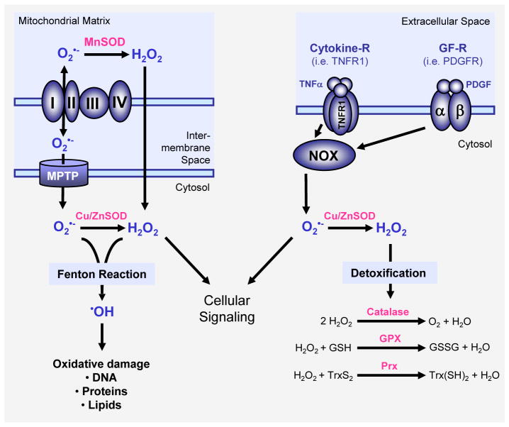
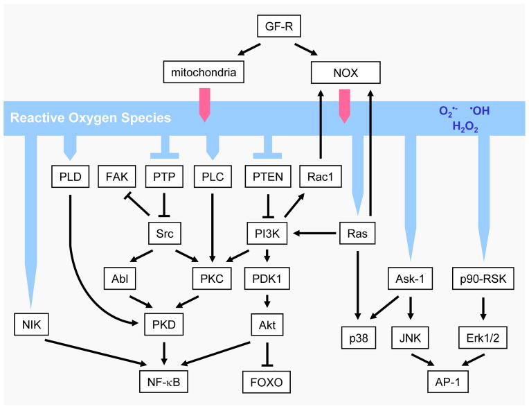
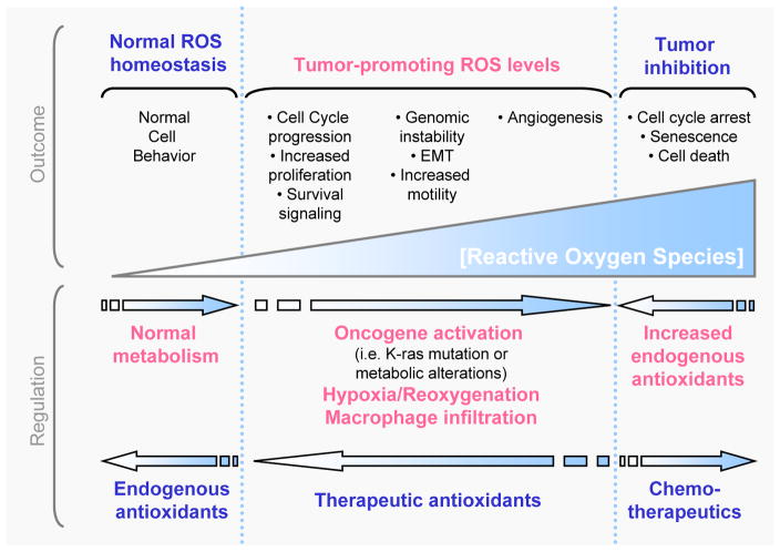
Elevated rates of reactive oxygen species (ROS) have been detected in almost all cancers, where they promote many aspects of tumour development and progression. However, tumour cells also express increased levels of antioxidant proteins to detoxify from ROS, suggesting that a delicate balance of intracellular ROS levels is required for cancer cell function. Further, the radical generated, the location of its generation, as well as the local concentration is important for the cellular functions of ROS in cancer. A challenge for novel therapeutic strategies will be the fine tuning of intracellular ROS signalling to effectively deprive cells from ROS-induced tumour promoting events, towards tipping the balance to ROS-induced apoptotic signalling. Alternatively, therapeutic antioxidants may prevent early events in tumour development, where ROS are important. However, to effectively target cancer cells specific ROS-sensing signalling pathways that mediate the diverse stress-regulated cellular functions need to be identified. This review discusses the generation of ROS within tumour cells, their detoxification, their cellular effects, as well as the major signalling cascades they utilize, but also provides an outlook on their modulation in therapeutics.


Conflict of interest statement

Declaration of interest:

The authors report no conflicts of interest. The authors alone are responsible for the content and writing of the review.

Figures

Figure 1
Figure 1. Major mechanisms of ROS generation and detoxification
Superoxide (O2•−) radicals are generated at the inner membrane of the mitochondria as a byproduct of the electron transport chain and then release into the mitochondrial matrix or the cytosol via the mitochondrial permeability transition pore (MPTP). Superoxide is also generated through activation of NADPH oxidases (NOX) for example in response to growth factor receptor (GF-R) or cytokine receptor activation. SOD enzymes, such as MnSOD in the mitochondrial matrix or Cu/ZnSOD in the cytosol reduce superoxide to H2O2. Several cytosolic antioxidant systems, including catalase, glutathione peroxidase (GPX) and peroxiredoxins (Prx) detoxify cells from hydrogen peroxide by reducing it to water. Both hydrogen peroxide and superoxide contribute to cellular signaling but also can form hydroxyl radicals (OH). Hydroxyl radicals are generated from O2•− and H2O2 in the Fenton reaction and have damaging functions for proteins, DNA and lipids.
Figure 2
Figure 2. ROS-induced cellular signaling
Reactive oxygen species in cells can be generated by growth factor signaling through activation of the NADPH oxidase NOX1 or through the mitochondria. These ROS then can induce cellular signaling cascades by reversible oxidation of phosphatases such as PTEN or PTP in their active site cysteins or by direct oxidation of kinases such as Src. This leads to the activation of several signaling cascades such as a Src/PKD1-dependent NF-κB activation mechanism, the MAPK (Erk1/2, p38 and JNK) signaling cascades, as well as the PI3K/Akt signaling pathway. Other mechanisms, by which ROS induce cellular signaling is through activation of redox-regulated transcription factors such as AP-1 or FOXO.
Figure 3
Figure 3. Generation, regulation and effects of cellular ROS
ROS are generated in normal cellular processes and cells express antioxidants to deplete intracellular levels of oxygen radicals. Tumorigenic events including oncogene activation (i.e. mutation of K-ras), metabolic alterations or macrophage infiltration or hypoxia/reoxygenation processes in tissues can increase intracellular ROS levels and promote tumor formation or progression. These tumor-promoting ROS levels can lead to cell cycle progression, increased proliferation and survival signaling, EMT, increased motility, genomic instability and increased angiogenesis and may be negatively-regulated by therapeutic antioxidants. Finally, excessive increase in intracellular ROS levels as mediated by chemotherapeutics, can induce cell cycle arrest, senescence or cell death of tumor cells, but may be repulsed by the tumor cells through an increase in the expression of endogenous antioxidants.

References

    1. Storz P. Reactive oxygen species in tumor progression. Front Biosci. 2005;10:1881–96. - PubMed
    1. Szatrowski TP, Nathan CF. Production of large amounts of hydrogen peroxide by human tumor cells. Cancer Res. 1991;51(3):794–8. - PubMed
    1. Babio BM. NADPH oxidase: an update. Blood. 1999;93(5):1464–76. - PubMed
    1. Ha D, Williams E, Cadenas E. Mitochondrial respiratory chain-dependent generation of superoxide anion and its release into the intermembrane space. Biochem J. 2001;353(Pt 2):411–6. - PMC - PubMed
    1. Crompto M. The mitochondrial permeability transition pore and its role in cell death. Biochem J. 1999;341 (Pt 2):233–49. - PMC - PubMed

Publication types

Substances

LinkOut - more resources