

Evolving molecular diagnostics for familial cardiomyopathies: at the heart of it all
- PMID: 20370590
- PMCID: PMC5022563
- DOI: 10.1586/erm.10.13
Evolving molecular diagnostics for familial cardiomyopathies: at the heart of it all
Abstract
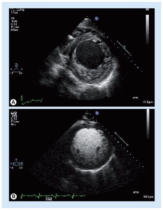
Cardiomyopathies are an important and heterogeneous group of common cardiac diseases. An increasing number of cardiomyopathies are now recognized to have familial forms, which result from single-gene mutations that render a Mendelian inheritance pattern, including hypertrophic cardiomyopathy, dilated cardiomyopathy, restrictive cardiomyopathy, arrhythmogenic right ventricular cardiomyopathy and left ventricular noncompaction cardiomyopathy. Recently, clinical genetic tests for familial cardiomyopathies have become available for clinicians evaluating and treating patients with these diseases, making it necessary to understand the current progress and challenges in cardiomyopathy genetics and diagnostics. In this review, we summarize the genetic basis of selected cardiomyopathies, describe the clinical utility of genetic testing for cardiomyopathies and outline the current challenges and emerging developments.
Figures
References
-
- Hershberger RE, Lindenfeld J, Mestroni L, et al. Genetic evaluation of cardiomyopathy – a Heart Failure Society of America practice guideline. J Card Fail. 2009;15(2):83–97. Comprehensively evaluated the evidence for genetic evaluation, clinical screening, and molecular genetic testing of cardiomyopathies (hypertrophic cardiomyopathy [HCM], dilated cardiomyopathy [DCM], restrictive cardiomyopathy [RCM], arrhythmogenic right ventricular cardiomyopathy [ARVC] and left ventricular noncompaction cardiomyopathy [LVNC]) based in published studies. - PubMed
-
- Maron BJ, Towbin JA, Thiene G, et al. Contemporary definitions and classification of the cardiomyopathies: an American Heart Association Scientific Statement from the Council on Clinical Cardiology, Heart Failure and Transplantation Committee; Quality of Care and Outcomes Research and Functional Genomics and Translational Biology Interdisciplinary Working Groups; and Council on Epidemiology and Prevention. Circulation. 2006;113(14):1807–1816. Presents the working framework for cardiomyopathies in the context of molecular genetics and their diverse phenotypes. Importantly, it recognizes the importance of molecular genetic testing and introduces several new entities, including LVNC, in the context of other cardiomyopathies. - PubMed
-
- Rodriguez JE, McCudden CR, Willis MS. Familial hypertrophic cardiomyopathy: basic concepts and future molecular diagnostics. Clin Biochem. 2009;42(9):755–765. - PubMed
-
- Sen-Chowdhry S, Syrris P, McKenna WJ. Role of genetic analysis in the management of patients with arrhythmogenic right ventricular dysplasia/cardiomyopathy. J Am Coll Cardiol. 2007;50(19):1813–1821. - PubMed
Website
-
- Medical genetics information resource. www.genetests.org.
Publication types
MeSH terms
Grants and funding
LinkOut - more resources
Full Text Sources
Medical