Review
. 2010 Jun;16(3):137-62.
doi: 10.1111/j.1755-5949.2010.00142.x. Epub 2010 Mar 29.

Acetylcholine-dopamine interactions in the pathophysiology and treatment of CNS disorders

Affiliations
Review

Acetylcholine-dopamine interactions in the pathophysiology and treatment of CNS disorders

Deranda B Lester et al. CNS Neurosci Ther. 2010 Jun.

Abstract

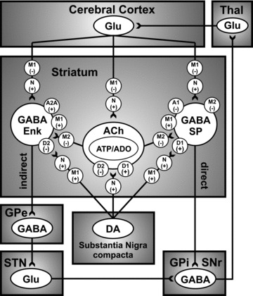
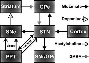
Dopaminergic neurons in the substantia nigra pars compacta and ventral tegmental area of the midbrain form the nigrostriatal and mesocorticolimbic dopaminergic pathways that, respectively, project to dorsal and ventral striatum (including prefrontal cortex). These midbrain dopaminergic nuclei and their respective forebrain and cortical target areas are well established as serving a critical role in mediating voluntary motor control, as evidenced in Parkinson's disease, and incentive-motivated behaviors and cognitive functions, as exhibited in drug addiction and schizophrenia, respectively. Although it cannot be disputed that excitatory and inhibitory amino acid-based neurotransmitters, such as glutamate and GABA, play a vital role in modulating activity of midbrain dopaminergic neurons, recent evidence suggests that acetylcholine may be as important in regulating dopaminergic transmission. Midbrain dopaminergic cell tonic and phasic activity is closely dependent upon projections from hindbrain pedunculopontine and the laterodorsal tegmental nuclei, which comprises the only known cholinergic inputs to these neurons. In close coordination with glutamatergic and GABAergic activity, these excitatory cholinergic projections activate nicotinic and muscarinic acetylcholine receptors within the substantia nigra and ventral tegmental area to modulate dopamine transmission in the dorsal/ventral striatum and prefrontal cortex. Additionally, acetylcholine-containing interneurons in the striatum also constitute an important neural substrate to provide further cholinergic modulation of forebrain striatal dopaminergic transmission. In this review, we examine neurological and psychopathological conditions associated with dysfunctions in the interaction of acetylcholine and dopamine and conventional and new pharmacological approaches to treat these disorders.


Conflict of interest statement

The authors have no conflict of interest.

Figures

Figure 1
Figure 1
Simplified thalamocortical basal ganglia circuitry depicting the innervation of the striatum by the nigrostriatal DA system and mediation of excitatory and inhibitory influence, via dopamine D1‐like (D1/D5) and D2‐like (D2/D3/D4) receptors and acetylcholine (ACh) muscarinic M1‐like (M1/3/5) and M2‐like (M2/4) receptors, of direct and indirect GABAergic striatal output pathways to the globus pallidus internus/substantia nigra reticulata (GPi/SNr), respectively. Note that the direct GABAergic striatal output pathway contains both M1 and M2 receptors, whereas the indirect pathway has primarily M1 receptors [29]. Presynaptic nicotinic receptors (N), of subtypesincluding α4β2*, α6β2*, and α4α6β2β3*, may also modulate striatal dopamine release, as well as glutamate release via presynaptic α7 nicotinic and M1 (likely M3) muscarinic receptors [30]. Nigrostriatal dopamine may also interact with striatal cholinergic interneurons, via dopamine D1‐ and D2‐like receptors, to mediate the co‐release of adenosine triphosphate (ATP) and adenosine (ADO) to act on A1 and A2A receptors on direct and indirect GABAergic striatal output pathways, respectively. GABA: γ‐aminobutyric acid; Glu: glutamate; GPe: globus pallidus externus; Enk: enkephalin; SP: substance P; STN: subthalamic nucleus; Thal: thalamus. “+” and “–” depicts the excitatory and inhibitory influence of each receptor subtype on the activity of the dopaminergic.
Figure 2
Figure 2
Simplified basal ganglia circuitry depicting direct innervation of dopaminergic cells in the substantia nigra compacta (SNc) by the cholinergic and glutamatergic neurons in the pedunculopontine tegmental nucleus (PPT) and indirect innervation of the SNc via glutamatergic neurons in the STN. SNr: substantia nigra reticulata; GPi: globus pallidus internus; GPe: globus pallidus externus. The direct GABAergic pathway from striatum to SNr/GPi has been omitted for clarity.
Figure 3
Figure 3
Schematic diagram of the mouse brain illustrating a typical setup for in vivo fixed potential amperometric recording of striatal dopamine release in mice (C57BL/6J mice, Jackson Labs.) evoked by electrical stimulation (20 pulses of 0.5 ms duration cathodal monophasic constant current pulses at 50 Hz applied every 30 sec at 800 μA). A carbon fiber microelectrode (Thornel Type P, Union Carbide) is positioned in the dorsomedial striatum, an Ag/AgCl reference and stainless‐steel auxiliary electrode combination is placed in contact with contralateral cortical tissue, a concentric bipolar stimulating electrode (SNE‐100; Rhodes Medical Co.) is implanted into the pedunculopontine tegmental nucleus (PPT), and a stainless‐steel guide cannula is placed in the STN for microinfusions of the axonal blocker lidocaine, nAChR, or mAChR antagonists. Coordinates (in mm) for the PPT, STN guide cannula, and striatum were (AP −4.7, −2.0, +1.4 from bregma, ML +1.25, +1.6, +1.4 from midline, and DV −2.7, −3.0, −2.5 from dura), respectively. In other studies examining STN stimulation and intra‐substantia nigra (SN) microinfusions described later, coordinates corresponded to (in mm) (AP −2.0, −3.1 from bregma, ML +1.6, +1.35 from midline, and DV −4.0, −2.8 from dura), respectively [55]. Black, light gray, and dark gray neuronal pathways correspond to glutamatergic, cholinergic, and dopaminergic neurons.
Figure 4
Figure 4
Mean amperometric recordings of dopamine release in the striatum evoked by electrical stimulation of the pedunculopontine tegmental nucleus (A) and corresponding mean peak percentages (B). Profiles illustrate mean peak effects in response to STN microinfusions of PBS, the local anaesthetic lidocaine, or a combination of the muscarinic acetylcholine receptor (mAChR) antagonist scopolamine (scop) and the nicotinic acetylcholine receptor (nAChR) antagonist mecamylamine (mec) (A). Time zero indicates the start of the train of 20 pulses at 50 Hz. *Significant change in striatal dopamine concentration after the infusion compared to pre‐infusion baseline responses (100%).
Figure 5
Figure 5
Mean amperometric recordings of dopamine release in the striatum evoked by electrical stimulation of the STN (A) and corresponding mean peak percentages (B). Profiles illustrate mean peak effects in response to substantia nigra pars compacta (SNc) microinfusions of PBS, the mAChR antagonist scopolamine, or the nAChR antagonist mecamylamine (A). Time zero indicates the start of the train of 20 pulses at 50 Hz. *Significant change in striatal dopamine concentration after the infusion compared to pre‐infusion baseline responses (100%).
Figure 6
Figure 6
Simplified mesocorticolimbic circuitry depicting dopaminergic (DA) cells in the VTA projecting to the core and shell region of the nucleus accumbens (NAc) and medial prefrontal cortex and excitatory (acetylcholine/glutamate) inputs from the laterodorsal tegmental nucleus (LDT) and associated excitatory (glutamate) and inhibitory (γ‐aminobutyric acid: GABA) feedback from cortex and VP.
Figure 7
Figure 7
Mean amperometric recordings of dopamine release in the nucleus accumbens evoked by electrical stimulation of the laterodorsal tegmental nucleus (A) and corresponding mean peak percentages in bar graph form (B). Profiles illustrate mean peak effects in response to VTA microinfusions of PBS, the mAChR antagonist scopolamine, or the nAChR antagonist mecamylamine (A). Time zero indicates the start of the train of 20 pulses at 50 Hz. *Significant change in striatal dopamine concentration after the infusion compared to pre‐infusion baseline responses (100%). Coordinates in mm for the stimulating electrode in the LDT, guide infusion cannula in the VTA, and carbon fiber recording microelectrode in the NAc were: AP −1.0, +0.9, +1.5 from lambda, ML +0.4, +0.3, +1.0 from midline, and DV −2.4, −4.0, −4.0 from dura), respectively [55].
Figure 8
Figure 8
Mean amperometric recordings of dopamine release in the nucleus accumbens evoked by electrical stimulation of the laterodorsal tegmental nucleus (LDT) (A) and time courses of the effects of scopolamine prior to (B) or following (C) cocaine administration. Profiles illustrate mean peak effects in response to VTA microinfusions of PBS or scopolamine (scop, 10 Jg/0.5 Jl) prior to or following intraperitoneal injection (IP) of saline (10 mL/kg) or cocaine (10 mg/kg), with respect to pre‐drug baseline responses (100%) (A). Timezero indicates the start of the train of 20 pulses at 50 Hz. *Significantly higher dopamine levels following cocaine compared to saline injection. #Significantly lower dopamine levels following scopolamine compared to PBS infusion.

Similar articles

Cited by

References

    1. Albanese A, Minciacchi D. Organization of the ascending projections from the ventral tegmental area: A multiple fluorescent retrograde tracer study in the rat. J Comp Neurol 1983;216:406–420. - PubMed
    1. Bjorklund A, Lindvall O. Dopamine‐containing systems in the CNS. In: Bjorklund A, Hokfelt T, editors. Handbook of chemical neuroanatomy. Classical transmitters in the CNS (Volume 2). Amsterdam : Elsevier, 1984;55–122.
    1. Fallon GH, Moore RY. Catecholamine innervation of the basal forebrain. IV. Topography of the dopamine projection to the basal forebrain and neostriatum. J Comp Neurol 1978;180:545–580. - PubMed
    1. Le Moal M, Simon H. Mesocorticolimbic dopaminergic network: Functional and regulatory roles. Physiol Rev 1991;71:155–234. - PubMed
    1. Jellinger KA. Recent developments in the pathology of Parkinson's disease. J Neural Transm Suppl 2002;62:347–376. - PubMed

MeSH terms