. 2010 Apr 2;141(1):142-53.
doi: 10.1016/j.cell.2010.02.023.

A global in vivo Drosophila RNAi screen identifies NOT3 as a conserved regulator of heart function

Affiliations

A global in vivo Drosophila RNAi screen identifies NOT3 as a conserved regulator of heart function

G Gregory Neely et al. Cell. .

Abstract

Heart diseases are the most common causes of morbidity and death in humans. Using cardiac-specific RNAi-silencing in Drosophila, we knocked down 7061 evolutionarily conserved genes under conditions of stress. We present a first global roadmap of pathways potentially playing conserved roles in the cardiovascular system. One critical pathway identified was the CCR4-Not complex implicated in transcriptional and posttranscriptional regulatory mechanisms. Silencing of CCR4-Not components in adult Drosophila resulted in myofibrillar disarray and dilated cardiomyopathy. Heterozygous not3 knockout mice showed spontaneous impairment of cardiac contractility and increased susceptibility to heart failure. These heart defects were reversed via inhibition of HDACs, suggesting a mechanistic link to epigenetic chromatin remodeling. In humans, we show that a common NOT3 SNP correlates with altered cardiac QT intervals, a known cause of potentially lethal ventricular tachyarrhythmias. Thus, our functional genome-wide screen in Drosophila can identify candidates that directly translate into conserved mammalian genes involved in heart function.


Figures

Figure 1
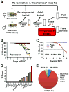
Figure 1. Genome wide screen for conserved heart genes
(A) Schematic for screen setup. TinΔ4-Gal4, a cardiac tissue specific driver, was used to drive conserved UAS-RNAi hairpins in the developing heart. Developmental lethality and baseline adult viability was scored. Viable adult flies were then given a heart stress (continued exposure to 29°C) and survival was scored on day 6. Fly lines showing a potential developmental or heart function phenotype were then retested to confirm the candidate gene. (B) 80 randomly selected UAS-RNAi lines were crossed to TinCΔ4-Gal4 and evaluated for adult lethality following an increase in ambient temperate as cardiac stressor. Lines were either viable (black) or died starting around day 3. Data from individual lines are shown as % survival on the indicated days. (C) Mean responses from viable and failing (death after exposure to 29°C) flies revealed an average lethal time at which 50% of failing flies died (LT50) of 6.19 days. (D) Efficacy of TinCΔ4-Gal4 x UAS-RNAi lines to knock-down transcription factors known to play a role in heart formation. (E) Using this system a genome-wide screen was performed to search for conserved candidate genes for adult heart function under conditions of cardiac stress. 4.6% TinCΔ4-Gal4 x UAS-RNAi lines were developmental lethal. Among the 7971 viable lines, 490 transformant lines exhibited significantly increased death (Z-score >3, determined on day 6 after shifting the ambient temperature to 29°C). See also Figure S1 and Tables S1 and S2.
Figure 2
Figure 2. A global network of heart function
The systems network includes data from the significantly enriched Drosophila KEGG and mouse and human KEGG and C2 data sets. Pathways and gene sets from the same biological processes were grouped into common functional categories. Green nodes represent statistically enriched functional categories of pathways; red nodes represent direct primary fly RNAi hits; light red nodes represent their first degree binding partners; and blue nodes indicate genes that were scored as developmentally lethal in our Drosophila heart screen. Lines indicate associations of the genes to the appropriate functional category. All KEGG pathways and selected C2 gene sets have been represented in the systems map. See also Tables S3–S5.
Figure 3
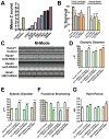
Figure 3. The CCR4-Not complex is a central regulator of adult heart function and loss of not3 results in dilated cardiomyopathy in Drosophila
(A) Mean Z scores for TinCΔ4-Gal4 x UAS-RNAi lines targeting the indicated members of the fly CCR4-Not complex. A negative control (w1118 [isogenic to the RNAi library] X TinΔ4-Gal4) and the positive control Tinman RNAi line are shown. (B) Not1, not3, and UBC4 are essential for proper adult heart function in both Tinman and Hand-expressing cells. Data are shown as mean +/− SEM for at least 3 replicates. RNAi1 and RNAi2 indicate different transgenic hairpins targeting not3. * p<0.05, ** p<0.01 by ANOVA. (C) 1 week old adult flies with Hand-Gal4 driving not3 or UBC4 cardiac specific knock-down exhibit impaired heart function. M-modes provide traces of the heart contractions to document the movements in a 1 pixel-wide region of the heart tube over time. HandG4/+ control are the progeny of Hand-Gal4 crossed to w1118. Hand>Not3-RNAi are the progeny of Hand-Gal4 crossed to either UAS-not3-RNAi (−1 or −2) or UAS-UBC4-RNAi lines. Fly heart analysis was generated using a MatLab based image analysis program (Fink et al., 2009; Ocorr et al., 2007b). M-modes of the RNAi knockdown hearts reveal dilated diastolic and systolic diameters (double-headed red arrows) and reduced shortening properties (difference between diameters) when compared to M-modes of control hearts. Each trace represents a 5 second recording. (D–G) Not3 or UBC4 heart specific knock-down perturbs several indices of cardiac performance. Progeny of Hand-Gal4 crossed to two different UAS-not3-RNAi lines or an UAS-UBC4-RNAi line (experimental), and w1118 crossed to UAS-RNAi or Hand-Gal4 driver (controls) were used for these experiments as in C. not3 and UBC4 knockdown led to significantly wider (D) diastolic and (E) systolic diameters, and as a result significantly depressed (F) fractional shortening in all experimental lines relative to controls. (G) not3 knockdown trended toward a slight lengthening in the heart period (time between consecutive diastolic intervals) while UBC4 knockdown led to a significant increase in heart period. Mean values ± SEM are shown for each group (n = 29–40). Unpaired t-tests were performed between each Hand-Gal4>UAS-RNAi and each corresponding UAS-RNAi/+ control (progeny of w1118 crossed to UAS-RNAi line). Additionally, one-way ANOVAs with Bonferroni multiple comparison tests revealed no significant differences between the HandG4/+ control and all UAS-RNAi/+ control lines, for any cardiac parameter measured. * p<0.05, ** p<0.01, *** p<0.001. See also Figure S3 and Video S1.
Figure 4
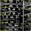
Figure 4. not3 and UBC4 cardiac specific RNAi-knockdown substantially perturb myofibrillar organization and content
(A) Alexa584-phalloidin staining of control Drosophila cardiac tubes reveals typical spiraling myofibrillar arrangements within the cardiomyocytes. The fibers, especially those in the conical chamber, located anteriorly, are densely packed with f-actin. (B–D) Relative to control hearts, not3 or UBC4 RNAi knockdown severely disrupts myofibrillar organization and leads to an apparent loss of myofilaments as noted by large gaps in f-actin staining (*) as well as by a lack of myosin heavy chain transcripts (Fig. S4F). (A-D′) Enlarged images of the conical chambers from A–D, respectively, which illustrate the high degree of myofibrillar disarray and large gaps in f-actin staining within the cardiomyocytes of not3 and UBC4 RNAi knockdown hearts. Original images taken at 10X magnification with a Zeiss Imager Z1 fluorescent microscope.
Figure 5
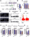
Figure 5. not3+/− mice exhibit reduced heart contractility, ex vivo function, and histone modifications that can be rescued by treatment with HDAC inhibitors
(A) Gene targeting strategy. Exons 2 to 9 of the not3 gene (official symbol cnot3) were replaced with a PGK-Neo cassette by homologous recombination in A9 ES cells. The wild type allele, the targeting vector, and mutant allele, and the PGK-Neo and DTH selection cassettes are shown. Blue boxes indicate exons. (B) Real-time PCR analyses for not3 mRNA expression in 3 month old wild-type and not3+/− hearts. Values were normalized to gapdh mRNA expression. n = 6 mice per group. (C) not3+/− mice display a significant reduction in % fractional shortening at 4 months of age, which became more pronounced with age. n = 6–8 mice per group. Fractional shortening was determined by echocardiography. (D) Representative M-mode echocardiography for wild-type and not3+/− mice at 8 months of age. (E) Left ventricular pressure (LVP) measurements in isolated ex vivo not3+/− and not3+/+ hearts under isoproterenol perfusion. not3+/− hearts from 4 months old mice showed impaired contractile responses to different doses of isoproterenol perfusion in the retrograde Langendorff mode as compared to age matched controls. (F) Impaired contractile response of ex vivo not3+/− hearts to electrical field stimulation (EFS) compared with littermate not3+/+ hearts. Representative data for left ventricular pressure (LVP) at 20V stimulation are shown. (G) H3K9 acetylation (H3K9ac), (H) H3K4 trimethylation (H3K4me3) and (I) H3K27 trimethylation (H3K27me3) levels were analyzed by Western blot for acid-extracted histones from whole heart ventricles of wild type and not3+/− mice treated with vehicle or VPA (0.71% w/v in drinking water for 1 week) Band intensities were normalized to total H3 levels. (J and K) Treatment (1 week) with the HDAC inhibitor VPA rescue impaired ex vivo heart contractility of not3+/− hearts to isoproterenol (100nM) perfusion or 25V EFS. All values are mean +/− SEM. *; P < 0.05, **; P < 0.01. n = 5–12 per group. See also Figure S4 and S5.
Figure 6
Figure 6. Not3+/− mice exhibit severe heart failure in response to pressure overload
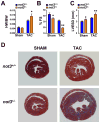
(A) Heart weight to body weight ratios (HW/BW) in not3+/− and not3+/+ littermate mice 3 weeks after transverse aortic constriction (TAC). Animals receiving sham surgery are shown as controls. (B) and (C) Echocardiography of male not3+/− and wild type littermates 3 weeks after TAC. not3+/− mice with TAC show (B) decreased % fractional shortening (%FS) and (C) increased left ventricular diameter in systolic phase (LVESD) compared with not3+/+ mice that received TAC. (D) Representative sections of not3+/+ and not3+/− hearts analyzed 3 weeks after sham or TAC surgery. Masson-trichrome staining are shown to visualize collagen deposits indicative of fibrotic changes. Note the severe cardiac hypertrophy and ventricular dilation in not3+/− mice following TAC.
Figure 7
Figure 7. Not3 is a conserved regulator of heart function
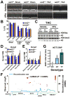
(A and B) Rescue of severe heart failure in TAC not3+/− hearts by the HDAC inhibitor VPA. One day after TAC or sham surgery the mice received treatment with vehicle or VPA (0.71% w/v in drinking water) for 6 weeks. (A) Representative M-mode echocardiography and (B) % FS in not3+/− and not3+/+ littermate mice 6 weeks after TAC or sham surgery with or without VPA treatment. (C) Reduced H3K9 acetylation (H3K9ac) and H3K4 trimethylation (H3K4me3) levels were rescued by VPA treatment. Acid-extracted histones from the hearts 6 weeks after TAC surgery were immunoblotted with antibodies for H3K9ac and H3K4me3. H3 is shown as a loading control. (D and E) Real time PCR analyses for the QT interval-associated potassium channel genes Kcnq1 and Kcne1. Total RNA was isolated from hearts 6 weeks after TAC or sham surgery with or without VPA treatment, and Kcnq1 and Kcne1 mRNA levels were measured and normalized to 18S mRNA. Data are shown as fold changes compared to not3+/+ mice for each treatment group. Values are mean +/− SEM. *; P < 0.05, **; P < 0.01. n = 5–10 per group. (F) Regional visualization of the association signal between common variants in the NOT3 region and the adjusted QT interval (QTc). SNP rs36643 in the 5′ region of NOT3 (−969bp from the transcription start and −924 from the TATA box) showed a significant regional association (p=0.000366). (G) Association between the T allele of SNP rs36643 and a prolongation of QTc. * P< 0.0005 from linear regression with inverse variance weighting using an additive genetic model. Data is derived from a meta-analysis of genome-wide association scans in several populations (Pfeufer et al., 2009). See also Figure S6.

Similar articles

Cited by

References

    1. A.H.A. 2005. http://www.americanheart.org/
    1. Alayari NN, Vogler G, Taghli-Lamallem O, Ocorr K, Bodmer R, Cammarato A. Fluorescent Labeling of Drosophila Heart Structures. J Vis Exp 2009 - PMC - PubMed
    1. Albert TK, Lemaire M, van Berkum NL, Gentz R, Collart MA, Timmers HT. Isolation and characterization of human orthologs of yeast CCR4-NOT complex subunits. Nucleic acids research. 2000;28:809–817. - PMC - PubMed
    1. Behm-Ansmant I, Rehwinkel J, Doerks T, Stark A, Bork P, Izaurralde E. mRNA degradation by miRNAs and GW182 requires both CCR4:NOT deadenylase and DCP1:DCP2 decapping complexes. Genes & development. 2006;20:1885–1898. - PMC - PubMed
    1. Bodmer R. Heart development in Drosophila and its relationship to vertebrates Trends in cardiovascular medicine. 1995;5:21–28. - PubMed

Publication types