. 2010 Jan 27;2(16):16ra7.
doi: 10.1126/scitranslmed.3000389.

Resiliency and vulnerability in the HER2-HER3 tumorigenic driver

Affiliations

Resiliency and vulnerability in the HER2-HER3 tumorigenic driver

Dhara N Amin et al. Sci Transl Med. .

Abstract

About 25% of breast cancers harbor the amplified oncogene human epidermal growth factor receptor 2 (HER2) and are dependent on HER2 kinase function, identifying HER2 as a vulnerable target for therapy. However, HER2-HER3 signaling is buffered so that it is protected against a nearly two-log inhibition of HER2 catalytic activity; this buffering is driven by the negative regulation of HER3 by Akt. We have now further characterized HER2-HER3 signaling activity and have shown that the compensatory buffering prevents apoptotic tumor cell death from occurring as a result of the combined loss of mitogen-activated protein kinase (MAPK) and Akt signaling. To overcome the cancer cells' compensatory mechanisms, we coadministered a phosphoinositide 3-kinase-mammalian target of rapamycin inhibitor and a HER2 tyrosine kinase inhibitor (TKI). This treatment strategy proved equivocal because it induced both TKI-sensitizing and TKI-desensitizing effects and robust cross-compensation of MAPK and Akt signaling pathways. Noting that HER2-HER3 activity was completely inhibited by higher, fully inactivating doses of TKI, we then attempted to overcome the cells' compensatory buffering with this higher dose. This treatment crippled all downstream signaling and induced tumor apoptosis. Although such high doses of TKI are toxic in vivo when given continuously, we found that intermittent doses of TKI administered to mice produced sequential cycles of tumor apoptosis and ultimately complete tumor regression in mouse models, with little toxicity. This strategy for inactivation of HER2-HER3 tumorigenic activity is proposed for clinical testing.


Figures
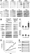
Fig. 1
Fig. 1. The durability and dose-dependency of lapatinib effects on HER signaling
A) SkBr3 cells were treated with the indicated concentrations of lapatinib for the indicated lengths of time (in hours). For treatments longer than 24 hours, the media was replaced with fresh media containing fresh drug every 24 hours. Total cell lysates were immunoblotted as indicated. B) SkBr3 cells were treated with the indicated concentrations of lapatinib for one hour or for 48 hours. Cell lysates were immunoblotted as indicated. C) SkBr3 cells were treated for the indicated times with 200nM lapatinib, and the relative expression of HER2 and HER3 mRNA was evaluated by real-time RT-PCR analysis of total cellular RNA with HER2 and HER3-specific primers and normalization with β-microglobulin. Results are expressed as fold change and are the average of triplicates. Error bars represent SEM. *, significant induction compared with 0 hours (p=0.004). D) SkBr3 cells were placed in media containing 50nM lapatinib or vehicle and the number of cells counted daily. The media was replaced every 24 hours. The data are shown as a percentage of the starting cell count. Results are the average of triplicates and error bars represent SEM E) SkBr3 cells were treated with 200nM or 1uM lapatinib for 48 hours. Cell lysates were assayed with a phospho-RTK antibody array identifying activated receptor tyrosine kinases. Each RTK is spotted in duplicate on a membrane, and the location of the duplicate EGFR, HER2, HER3, and HER4 spots, in this order, are indicated by X in the index. The exposures have been normalized such that the positive control spots in the four corners have identical intensities.
Fig. 2
Fig. 2. Signal buffering capacity in the HER2-HER3 dimer
A) Schematic diagram depicting how the transient inhibition of HER2-HER3 signaling and its sustained inhibition exhibit different sensitivities to lapatinib. The two-log difference in the minimal inhibitory concentration required for full inactivation (MIC100) reflects a two-log signal buffering capacity in HER2-HER3 signaling. B) SkBr3 cells were treated with the indicated concentrations of erlotinib for one hour or for 48 hours. Cell lysates were immunoblotted as indicated.
Fig. 3
Fig. 3. Akt underlies HER3 signal buffering and protection against lapatinib
A) SkBr3/ myrAktΔER and SkBr3/ myr*AktΔER cells were treated with 4-hydroxy tamoxifen (4HT) to induce the activation of the engineered Akt construct and lapatinib at 0.2 or 5 μM for the indicated times. Cell lysates were immunoblotted as indicated. Arrows, endogenous cellular Akt; starred arrows, the larger engineered Akt-ΔER fusion proteins. B) SkBr3/ myr*AktΔER and SkBr3/ myrAktΔER cells were treated with 4HT to induce the activation of the engineered Akt construct and 200nM lapatinib for the indicated times (in hours). Cellular RNA was extracted and the relative expression of HER3 mRNA was assayed by real-time quantitative PCR with HER3 specific primers. GAPDH was used as normalization control. The results shown are the average of triplicates, and error bars represent SEM. Data are relative expression with values from untreated cells set as 1. * indicates significantly different from the adjacent induced data point (p=0.005), and significantly different from the 0 timepoint (p=0.01).
Fig. 4
Fig. 4. Feedback and cross-talk in the MAPK and Akt pathways
A) SkBr3 cells were treated with the indicated concentrations of the specified PI3K inhibitors for the indicated times. Cell lysates were immunoblotted as indicated. B) SkBr3 cells were treated with 5 uM U0126 for the indicated durations of time. Cell lysates were immunoblotted as indicated. C) SkBr3 cells were treated with the indicated drugs for 72 hours, and the fraction of apoptotic cells quantified by FACS analysis of DNA degradation. * (p=0.0003) and ** (p=0.0006) indicate the significant induction of apoptosis compared with the mock treatment arm. BE, BEZ235; LY, LY294002; W, Wortmannin
Fig. 5
Fig. 5. Effects of lapatinib and BEZ235 alone and in combination on signaling
A) SkBr3 cells were treated with 200nM lapatinib or two concentrations of BEZ235 as single agents or in combination for the indicated durations of time. Cell lysates were immunoblotted as indicated. B) SkBr3/myrAktΔER and SkBr3/ myr*AktΔER cells were pre-treated with 4-hydroxy tamoxifen followed by treatment with the indicated concentrations of lapatinib or BEZ235 as single agents or in combination for 72 hours, and the fraction of apoptotic cells quantified by FACS analysis of DNA degradation. Significant protection from apoptosis by Akt induction is shown by * (p=0.00005) and ** (p=0.05). BE, BEZ235; LY, LY294002.
Fig. 6
Fig. 6. Inactivation of HER2-HER3 signaling by high dose lapatinib
A) HCC1569 cells were orthotopically grown in the mammary fat pad of nude mice. When the average tumor sizes reached approximately 100 mm3, mice were randomized to three groups of 10 mice each. One group was treated with lapatinib at 100mg/kg/day every day for 42 days. This is the maximum tolerated dose of lapatinib in continuous dosing. A second group was treated with lapatinib at 800mg/kg/day for a 5-day cycle followed by a 9-day off-treatment period for a total of three cycles. This is the maximum tolerated dose of lapatinib in this intermittent schedule. All treatments were administered by oral gavage in divided twice-daily dosing. Mice in the control arm were treated daily with vehicle. Tumor measurements were taken once or twice weekly. The treatment days for the continuous and high-dose intermittent arms are indicated below the graphs. Results shown are averages with the error bars indicating SEM. B) After completion of the 42-day treatment period, these mice were followed for another 30 days and tumor re-growth was measured and followed. Results shown are averages with the error bars indicating SEM. C) HCC1569 tumors from mice treated with the vehicle, 100mg/kg/day (low dose), or 800mg/kg/day (high dose) were resected 4 hours after the morning dose of the 5th day of treatment and lysates prepared from frozen tissues. The lysates were subjected to immunoblotting as indicated. D) An additional seven mice bearing HCC1569 tumors were treated with vehicle or lapatinib at 100 mg/kg/day or 800 mg/kg/day and plasma lapatinib concentrations were assessed 4 hours after the morning dose of the 5th day. Results represent average of replicates.

References

    1. Slamon DJ, Clark GM, Wong SG, Levin WJ, Ullrich A, McGuire WL. Human breast cancer: correlation of relapse and survival with amplification of the HER-2/neu oncogene. Science. 1987;235:177. - PubMed
    1. Ursini-Siegel J, Schade B, Cardiff RD, Muller WJ. Insights from transgenic mouse models of ERBB2-induced breast cancer. Nat Rev Cancer. 2007 May;7:389. - PubMed
    1. Moasser MM. The oncogene HER2: its signaling and transforming functions and its role in human cancer pathogenesis. Oncogene. 2007;26:6469. - PMC - PubMed
    1. Moody SE, Sarkisian CJ, Hahn KT, Gunther EJ, Pickup S, Dugan KD, Innocent N, Cardiff RD, Schnall MD, Chodosh LA. Conditional activation of Neu in the mammary epithelium of transgenic mice results in reversible pulmonary metastasis. Cancer Cell. 2002 Dec;2:451. - PubMed
    1. Blackwell KL, Pegram MD, Tan-Chiu E, Schwartzberg LS, Arbushites MC, Maltzman JD, Forster JK, Rubin SD, Stein SH, Burstein HJ. Single-agent lapatinib for HER2-overexpressing advanced or metastatic breast cancer that progressed on first- or second-line trastuzumab-containing regimens. Ann Oncol. 2009 Jun;20:1026. - PubMed

Publication types

MeSH terms