. 2010 Feb 3;2(17):17ra10.
doi: 10.1126/scitranslmed.3000116.

Inducing CTLA-4-dependent immune regulation by selective CD28 blockade promotes regulatory T cells in organ transplantation

Affiliations

Inducing CTLA-4-dependent immune regulation by selective CD28 blockade promotes regulatory T cells in organ transplantation

Nicolas Poirier et al. Sci Transl Med. .

Abstract

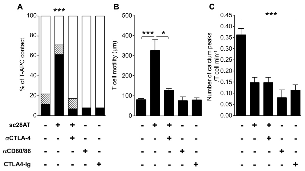
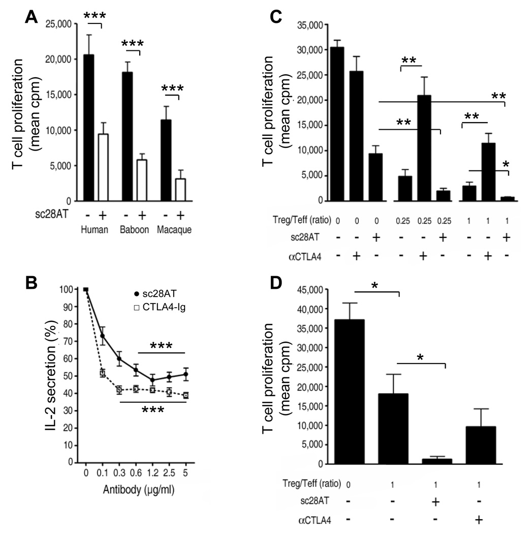
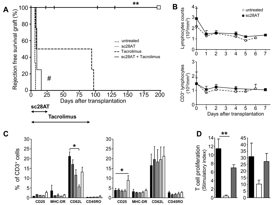
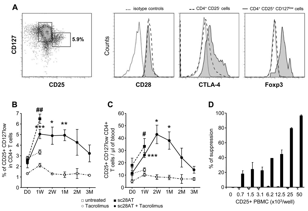
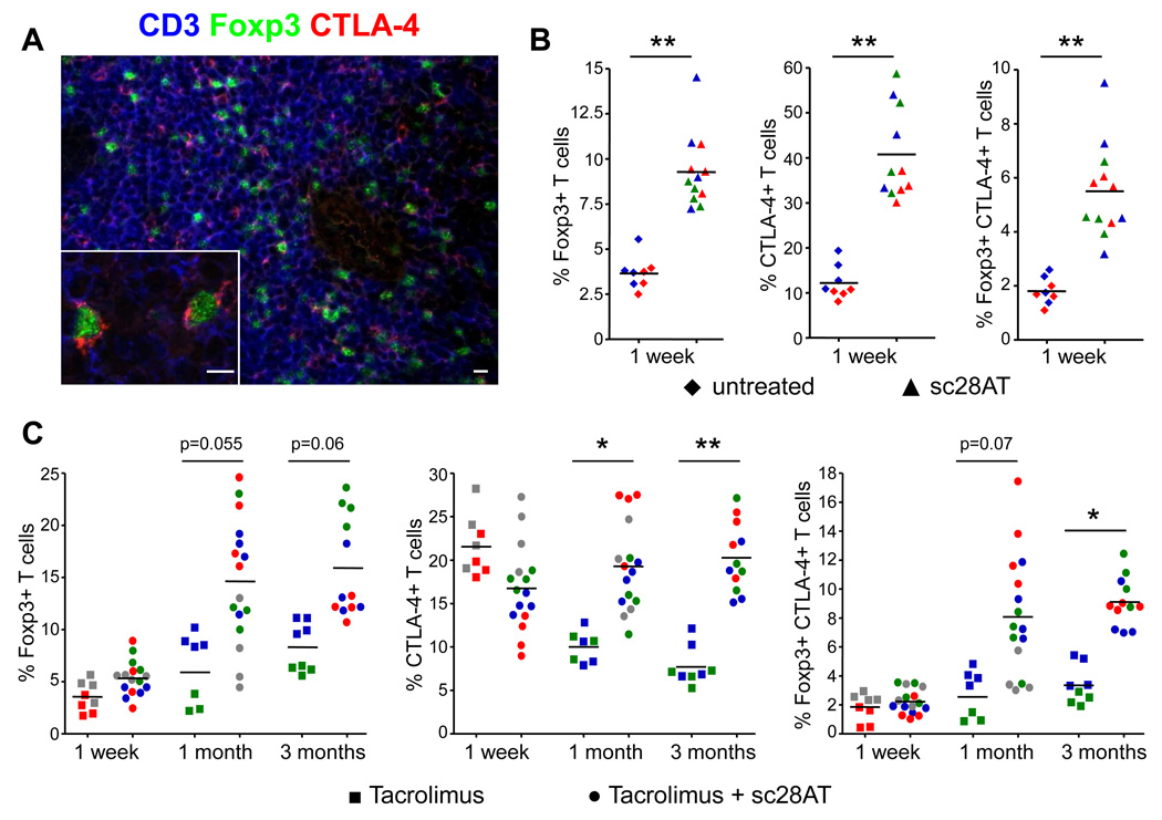
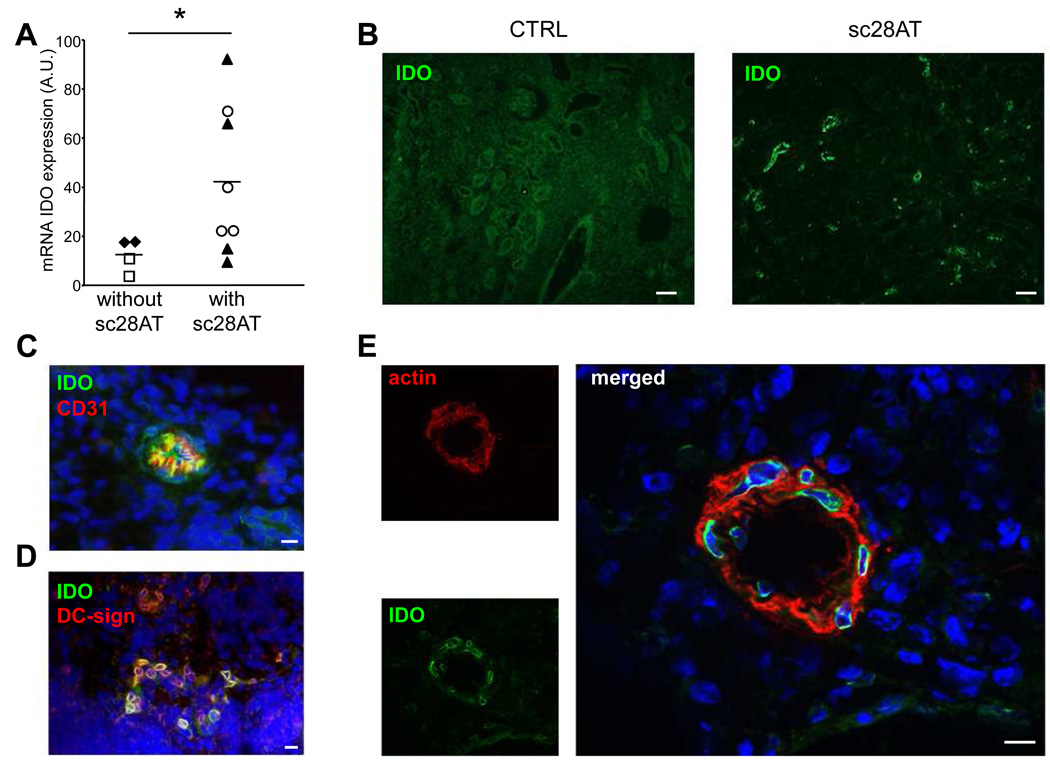
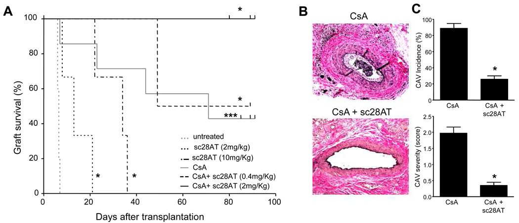
Transplantation is the treatment of choice for patients with end-stage organ failure. Its success is limited by side effects of immunosuppressive drugs, such as inhibitors of the calcineurin pathway that prevent rejection by reducing synthesis of interleukin-2 by T cells. Moreover, none of the existing drugs efficiently prevent the eventual rejection of the organ. Blocking the CD28-mediated T cell costimulation pathway is a nontoxic alternative immunosuppression strategy that is now achieved by blockade of CD80/86, the receptor for CD28 on antigen-presenting cells. However, interaction of CD80/86 with cytotoxic T lymphocyte-associated antigen 4 (CTLA-4) is required for immune regulation. Therefore, CD28 blockade, instead of CD80/86 blockade, might preserve regulatory signals mediated by CTLA-4 and preserve immune regulation. By using monovalent antibodies, we identified true CD28 antagonists that induced CTLA-4-dependent decreased T cell function compatible with regulatory T (Treg) cell suppression. In transplantation experiments in primates, blocking CD28 augmented intragraft and peripheral blood Treg cells, induced molecular signatures of immune regulation, and prevented graft rejection and vasculopathy in synergy with calcineurin inhibition. These findings suggest that targeting costimulation blockade at CD28 preserves CTLA-4-dependent immune regulation and promotes allograft survival.


Figures

Figure 1
Figure 1. CTLA-4-dependent impairment of T-APC contact after selective CD28 blockade
(A) Time-lapse microscopy of cognate contacts between EBV-specific T cells and B-EBV APCs in the presence of the indicated Ab. Results are shown as % of cells establishing short (≤ 5 min.; black), medium (between 5 and 15 min., hatched) and long (≥ 15 min., white) contacts over 20 min. (B) T cells were cultured as in (A) and the cumulative distance moved of individual cells was recorded to assess T cell motility. (C) T cells were cultured as in (A) and calcium peaks were recorded. * and *** indicate a significant difference at P<0.05 and 0.001, respectively.
Figure 2
Figure 2. Monovalent CD28 antagonists block allorecognition but do not impede the function of Treg cells in vitro
(A) Mixed-lymphocyte reaction using human (n=10), baboon (n=15) or macaque (n=9) PBMC. Black bars: mean ±SD in control conditions (mouse irrelevant IgG1). White bars: mean ± SD with 10 µg/ml sc28AT. (B) IL-2 secretion by Jurkat T cells stimulated with bacterial superantigen (staphylococcal enterotoxin E, SEE) and Raji B cells in the presence of sc28AT (n=8) or CTLA4-Ig (n=3). Results are expressed as percentage of IL-2 secretion observed in the absence of Ab (100%). (C) Suppressive activity of human Treg is not impeded by CD28 blockade. Tregs were added to CD4+CD25 T cells stimulated with allogeneic irradiated PBMC at the indicated ratio in the presence of 10µg/ml of CD28 or CTLA-4 blocking antibodies. Results are mean cpm ± SD of one representative assay out of 3. (D) Suppressive activity of human Treg pre-treated with CD28 or CTLA-4 blocking Ab. Treg were first cultured with allogeneic mature DC in the presence of sc28AT or anti-CTLA-4 Fab fragments (10µg/ml) for 18h, washed and assessed in a suppression assay. Results are mean cpm ± SD of a representative assay out of 3. *, ** and *** indicate a significant difference at p<0.05, 0.01 and 0.001, respectively.
Figure 3
Figure 3. Administration of sc28AT and Tacrolimus prevents kidney allograft rejection in baboons
(A) Rejection-free survival after renal allotransplantation for baboons without therapy (n=3) or treated with a 25 day induction therapy with sc28AT alone (n=4), Tacrolimus for 90 days (n=4) and sc28AT + Tacrolimus (25 and 90 days, respectively; n=5). Vertical hash marks, death of recipients without graft rejection as assessed by histology. White square, remaining living baboon. **, P=0.008 versus Tacrolimus monotherapy. ##, P=0.01 versus untreated controls. (B) Total lymphocyte counts (means ± SD, upper panel) and CD3+ T cell counts (lower panel) in untreated recipients (n=3) and recipients treated with sc28AT monotherapy (n=4). (C) Phenotype of blood T cells during the first week post transplantation in controls (n=3; left panel) and sc28AT monotherapy recipients (n=4; right panel). Percentages of T cells expressing the specified marker on day 0, 1, 2, 4 and 6 from left (black) to right (white). *, P<0.05. (D) Donor-specific hyporesponsiveness. PBMC were harvested from baboons in the sc28AT + Tacrolimus group and tested in MLR against donor cells before transplantation (black bars) and at day 90 (white bars) and against 3rd party cells at day 90 (grey bars). Results are mean stimulatory index ± SD of triplicate wells. Two experiments are shown. **, P=0.0014.
Figure 4
Figure 4. Treg cell enrichment in the peripheral blood of kidney allograft baboon recipients treated with sc28AT
(A) CD25+ CD127 lo Treg cells analyzed in blood by flow cytometry after gating on CD3+ CD4+ cells. These cells also expressed CD28, and intra-cellular CTLA-4 and Foxp3. (B-C) Kinetics of CD4+CD25+Foxp3+CD127lo Treg levels in blood, percentage of CD4+ T cells (B) or absolute number (C), in control untreated animals (n=3), sc28AT monotherapy (n=4), Tacrolimus monotherapy (n=4 until week 1 and then n=2), sc28AT + Tacrolimus (n=5 up to 2 weeks, 4 at 1 month and then n=3). D: days, W: weeks, M: month. # and ##, significant difference with the untreated group (P=0.02 and P=0.0011, respectively). ***, ** and * significant difference with the Tacrolimus monotherapy group (P<0.05, 0.01 and 0.001, respectively). (D) Suppressive activity of CD25+ PBMC from sc28AT + Tacrolimus recipients at day 90 after transplantation (black bars, n=3) or from control ungrafted baboons treated with Tacrolimus (white bars, n=4).
Figure 5
Figure 5. Increased Treg cell infiltration in kidney allografts following selective CD28 blockade with sc28AT
(A) Confocal-like microscopy analysis of a kidney graft biopsy from a sc28AT + Tacrolimus treated recipient one month post transplantation. Blue, CD3 staining; green, Foxp3 staining; red, CTLA-4 staining. Scale bars, 10µm. (B) Quantitative evaluation of graft infiltration by T cells expressing Foxp3, CTLA-4 or both in control and sc28AT monotherapy recipients 1 week post transplant (expressed as % of CD3+ T cells). Data from individual animals are represented with a specific color and the 4 individual data points represent quadruplicate evaluations performed on two different tissue sections of the same animal. (C) Same as (B) in recipients treated with Tacrolimus alone or sc28AT + Tacrolimus at 1 week, 1 month and 3 months post-transplant. * and ** indicate a significant difference at P<0.05 and 0.01, respectively.
Figure 6
Figure 6. Expression of indoleamine 2,3-dioxigenase (IDO) in allograft of sc28AT-treated recipients
(A) qPCR measurement of IDO mRNA transcripts one week post-transplantation in kidney graft biopsies from control untreated (♦, n=2) or Tacrolimus alone-treated recipients (□, n=2), and from sc28AT alone (▲, n=4) or sc28AT + Tacrolimus recipients (○, n=4). Each point represents the mean of duplicate measurements. *, P=0.04 (B) Immunohistology of biopsies from a control and a sc28AT-treated recipient labeled with an anti-IDO antibody (green) one week post transplantation. Scale bar, 100 µm. (C) Co-staining of IDO (green) and CD31, an endothelial cell marker (red), showing a small blood vessel. (D) Co-staining of IDO (green) and DC-sign, a C-type lectin expressed by immature myeloid cells (red), in the renal parenchyma. (E) Co-staining of IDO (green) and smooth muscle actin (red), showing co-localization in an arteriole. Nuclei are in blue. Scale bars in C, D and E, 10µm.
Figure 7
Figure 7. Selective CD28 inhibition prolongs cardiac allograft survival and prevents cardiac allograft vasculopathy in macaques
(A) Cardiac allograft survival for monkeys without therapy (n=5) or treated with sc28AT monotherapy at 2 mg/kg/day (n=3) or 10 mg/kg/day (n=3), Cyclosporine A monotherapy (n=6), Cyclosporine A + sc28AT at 0.4 mg/kg bitherapy (n=2) or Cyclosporine A + sc28AT at 2mg/kg bitherapy (n=3). *, P<0.05 and ***, P<0.001 (indicated group versus control untreated recipients). (B) A representative vessel from a cardiac allograft treated with Cyclosporine A (day 72, left panel) shows grade 2 cardiac allograft vasculopathy (CAV) with distinct neointimal thickening and 10–50% (estimated at 25% in this instance) luminal narrowing. In contrast, a representative graft artery from a recipient treated with sc28AT + Cyclosporine A shows an absence of neointimal proliferation (day 80, lower panel). (C) CAV incidence and severity at day 70–90, graded as described in Methods, were significantly lower (P<0.05) in CD28 blockade plus Cyclosporine A combination therapy (n=4) as compared with Cyclosporine A alone (n=5).

References

    1. Alegre ML, Frauwirth KA, Thompson CB. T-cell regulation by CD28 and CTLA-4. Nat Rev Immunol. 2001;1:220–228. - PubMed
    1. Kremer JM, Westhovens R, Leon M, Di Giorgio E, Alten R, Steinfeld S, Russell A, Dougados M, Emery P, Nuamah IF, Williams GR, Becker JC, Hagerty DT, Moreland LW. Treatment of rheumatoid arthritis by selective inhibition of T-cell activation with fusion protein CTLA4Ig. N Engl J Med. 2003;349:1907–1915. - PubMed
    1. Vincenti F, Larsen C, Durrbach A, Wekerle T, Nashan B, Blancho G, Lang P, Grinyo J, Halloran PF, Solez K, Hagerty D, Levy E, Zhou W, Natarajan K, Charpentier B. Costimulation blockade with belatacept in renal transplantation. N Engl J Med. 2005;353:770–781. - PubMed
    1. Takahashi T, Kuniyasu Y, Toda M, Sakaguchi N, Itoh M, Iwata M, Shimizu J, Sakaguchi S. Immunologic self-tolerance maintained by CD25+CD4+ naturally anergic and suppressive T cells: induction of autoimmune disease by breaking their anergic/suppressive state. Int Immunol. 1998;10:1969–1980. - PubMed
    1. Salomon B, Lenschow DJ, Rhee L, Ashourian N, Singh B, Sharpe A, Bluestone JA. B7/CD28 costimulation is essential for the homeostasis of the CD4+CD25+ immunoregulatory T cells that control autoimmune diabetes. Immunity. 2000;12:431–440. - PubMed

Publication types