. 2010 Feb 3;2(17):17ra9.
doi: 10.1126/scitranslmed.3000349.

Identification of therapeutic targets for quiescent, chemotherapy-resistant human leukemia stem cells

Affiliations

Identification of therapeutic targets for quiescent, chemotherapy-resistant human leukemia stem cells

Yoriko Saito et al. Sci Transl Med. .

Abstract

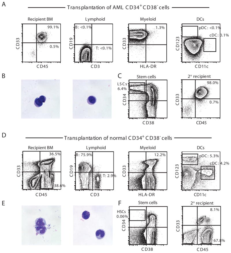
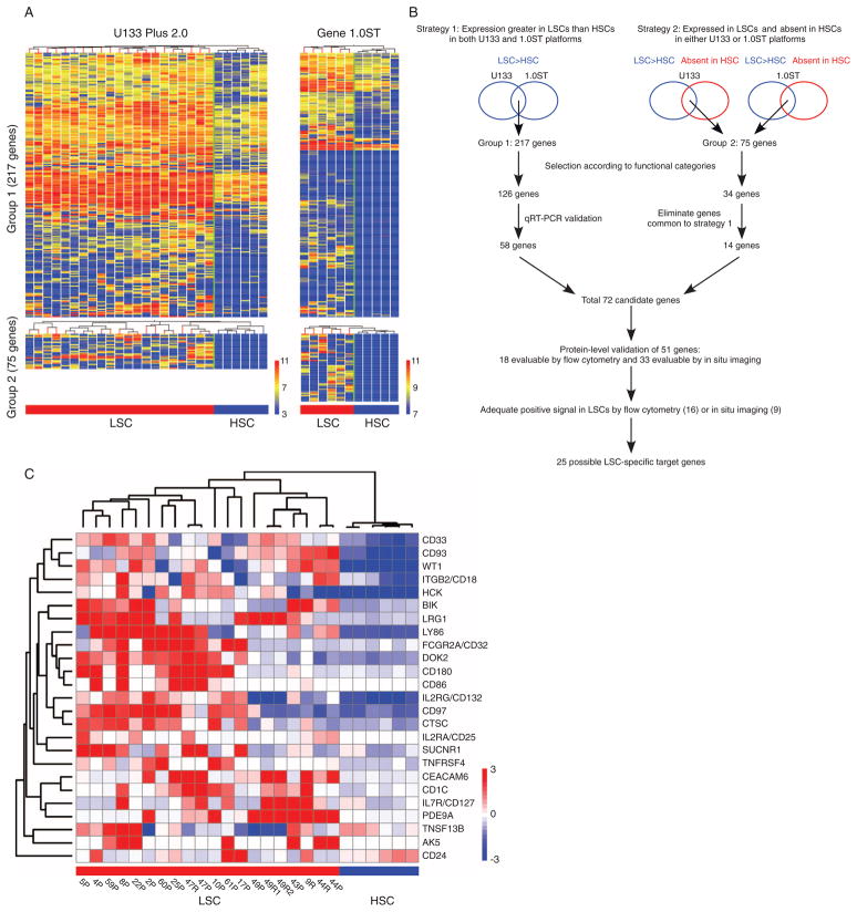
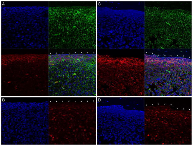
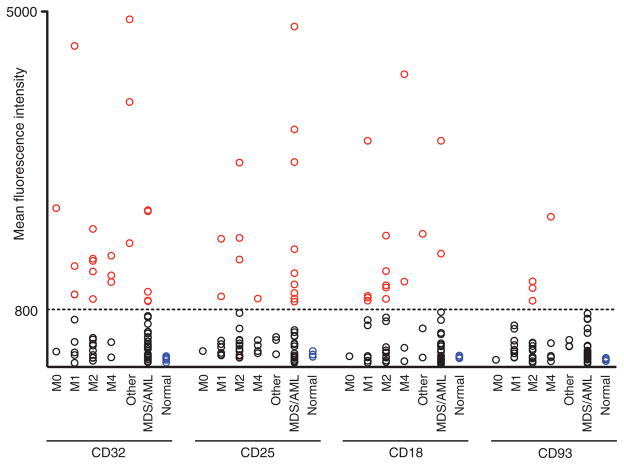
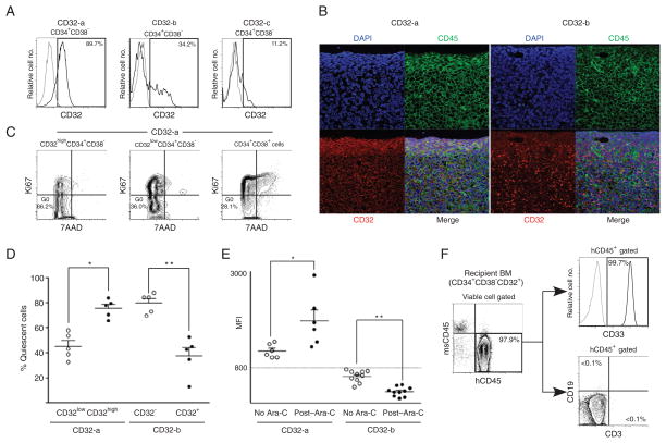
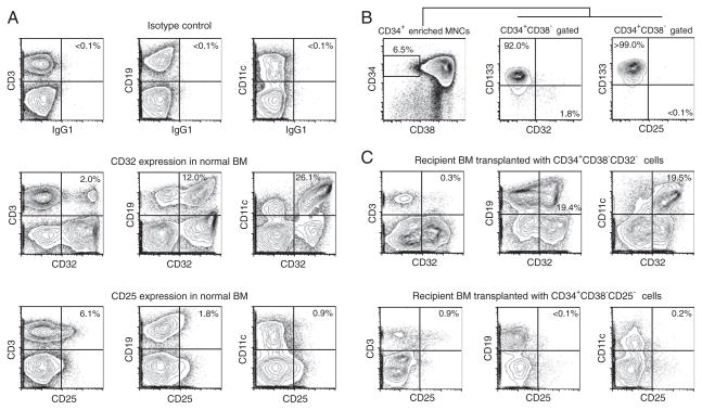
Human acute myeloid leukemia (AML) originates from rare leukemia stem cells (LSCs). Because these chemotherapy-resistant LSCs are thought to underlie disease relapse, effective therapeutic strategies specifically targeting these cells may be beneficial. Here, we report identification of a primary human LSC gene signature and functional characterization of human LSC-specific molecules in vivo in a mouse xenotransplantation model. In 32 of 61 (53%) patients with AML, either CD32 or CD25 or both were highly expressed in LSCs. CD32- or CD25-positive LSCs could initiate AML and were cell cycle-quiescent and chemotherapy-resistant in vivo. Normal human hematopoietic stem cells depleted of CD32- and CD25-positive cells maintained long-term multilineage hematopoietic reconstitution capacity in vivo, indicating the potential safety of treatments targeting these molecules. In addition to CD32 and CD25, quiescent LSCs within the bone marrow niche also expressed the transcription factor WT1 and the kinase HCK. These molecules are also promising targets for LSC-specific therapy.


Conflict of interest statement

Competing interests: The authors have no competing interests to declare.

Figures

Fig. 1
Fig. 1
CD34+CD38 phenotype functionally defines primary human AML LSCs and human normal HSCs in vivo. (A to C) Xenotransplantation of CD34+CD38 primary human AML cells results in initiation of AML. pDC, plasmacytoid DC; cDC, conventional DC. In AML-engrafted recipients, (A) BM is entirely replaced with hCD45+CD33+ cells with the complete absence of the normal immune subsets such as DCs, T cells and B cells and (B) PB hCD45+CD33+ cells show uniform-appearing blast-like morphology by May-Grunwald-Giemsa staining. (C) BM of recipients of CD34+CD38 AML cells was reconstituted with CD34+CD38, CD34+CD38+, and CD34 cells. Serial transplantation of CD34+CD38 cells results in complete replacement of the secondary recipient BM with hCD45+CD33+ cells. (D to F) Normal human CB CD34+CD38 cells are HSCs capable of long-term multilineage reconstitution of human hematopoiesis. In recipients of normal CB CD34+CD38 cells, (D) BM contains heterogeneous human hematopoietic cell types, including T cells, B cells, and DC subsets, by phenotype and (E) PB hCD45+CD33+ cells contain morphologically heterogeneous hematopoietic cells. (F) Transplantation of normal human CB CD34+CD38 cells results in the reconstitution of recipient BM with CD34+CD38, CD34+CD38+, and CD34 cells (left). BM of secondary CD34+CD38 cell recipient contains both hCD33+CD45+ human myeloid and hCD33 CD45+ human lymphoid cells (right).
Fig. 2
Fig. 2
LSC-specific gene candidates were derived from genes overrepresented in LSCs relative to HSCs. (A) Hierarchical clustering of genes overrepresented in AML LSCs relative to normal human HSCs identified by global expression pattern analyses with two microarray platforms. (B) Schematic representation of the analysis strategies integrating expression profiles obtained from two independent array platforms. (C) The expression of 25 candidate LSC-specific genes discriminate LSCs from HSCs. Patient ID and sample source (P, patient; R, recipient engrafted with the patient LSCs) are indicated below each column. The heat maps represent microarray signal intensity on a log2 scale.
Fig. 3
Fig. 3
LSC target molecules are expressed on cell cycle–quiescent cells in BM endosteal region. (A and C) WT1 (red)–positive hCD45 (green)–positive (A) and HCK (red)–positive hCD45 (green)–positive (C) AML cells are adjacent to the endosteum (*). (B and D) The cells adjacent to the endosteum (*) are also Ki67 (red)–negative. Nuclei are labeled with 4′,6-diamidino-2-phenylindole (DAPI) (blue). The BM contained >98% hCD45+CD33+ cells in each recipient. Scale bar, 20 μm.
Fig. 4
Fig. 4
Target molecules are present on the cell surface of primary AML LSCs. MFI of expression for CD32, CD25, CD18, and CD93 in 61 primary AML patient BM LSCs with normal human BM HSCs as controls. Cutoff MFI = 800 (indicated as a horizontal line). Red, black, and blue circles represent marker-positive AML LSCs, marker-negative AML LSCs, and normal BM HSCs, respectively. AML: M0, n = 2; M1, n = 8; M2, n = 15; M4, n = 4; other AML (M5, M7, and undetermined), n = 1 each; MDS/AML: n = 29; normal human BM: n = 5.
Fig. 5
Fig. 5
CD32 expression is associated with cell cycle quiescence and chemotherapy resistance of LSCs. (A) In CD32-positive AML, a single peak of CD32+ expression is present in LSCs (CD32-a, left panel). CD32-negative AML LSCs show two patterns of CD32 expression (CD32-b and CD32-c, middle and right panels). (B) In CD32-a AML-engrafted recipient (left), CD32 (red) is co-expressed with hCD45 (green) on AML cells abutting the BM endosteum (*). In CD32-b AML-engrafted recipient (right), there is scattered CD32 expression in the central region of the BM but no CD32 expression in the endosteal region. Nuclei are labeled with DAPI (blue). Scale bar, 20 μm. (C) Representative flow cytometry plots demonstrating enrichment of quiescent cells in CD32highCD34+CD38 population in CD32-positive AML. (D) In CD32-a LSCs, CD32-high LSCs are cell cycle–quiescent, whereas CD32-negative LSCs are cell cycle–quiescent in CD32-b LSCs (n = 5 for each group). *P = 0.0034, **P = 0.0006 by two-tailed t test. (E) CD32 expression in LSCs after in vivo Ara-C treatment. In vivo Ara-C treatment increases CD32 expression in CD32-positive (CD32-a) BM LSCs, indicating that CD32-low LSCs are preferentially eliminated by Ara-C (n = 6 each for control and Ara-C–treated group). *P = 0.0121. In contrast, CD32 expression decreases in CD32-negative (CD32-b) BM LSCs with Ara-C treatment (n = 10 each). **P < 0.0001 by two-tailed t test. (F) CD32+CD34+CD38 cells retain in vivo AML-initiating capacity after in vivo Ara-C treatment. Flow cytometry plots showing engraftment of hCD45+ cells in the BM of a representative recipient of purified CD34+CD38 CD32+ cells from an Ara-C–treated recipient. All hCD45+ human hematopoietic cells are also hCD33+ and there are no CD3+ T or CD19+ B cells present.
Fig. 6
Fig. 6
Depletion of LSC marker-positive cells does not affect normal human multilineage hematopoietic reconstitution in vivo. (A) CD32 and CD25 expression in normal human BM T, B, and myeloid cells with their corresponding isotypes. (B) In normal human CB, CD32+ and CD25+CD34+CD38 cells do not express CD133, a positive marker for normal HSCs. (C) Recipients of human normal CB depleted of CD32-positive cells are repopulated with both CD32-positive and CD32-negative human T, B, and myeloid cells. Similarly, human CB depleted of CD25-positive cells repopulated both CD25-positive and CD25-negative human T, B, and myeloid cells.

Similar articles

Cited by

References

    1. Horner MJ, Ries LAG, Krapcho M, Neyman N, Aminou R, Howlader N, Altekruse SF, Feuer EJ, Huang L, Mariotto A, Miller BA, Lewis DR, Eisner MP, Stinchcomb DG, Edwards BK. SEER Cancer Statistics Review 1975–2006. National Cancer Institute; Bethesda: 2009. [accessed 2 July 2009]. http://seer.cancer.gov/csr/1975_2006/, based on November 2008 SEER data submission.
    1. Breems DA, Van Putten WL, Huijgens PC, Ossenkoppele GJ, Verhoef GE, Verdonck LF, Vellenga E, De Greef GE, Jacky E, Van der Lelie J, Boogaerts MA, Löwenberg B. Prognostic index for adult patients with acute myeloid leukemia in first relapse. J Clin Oncol. 2005;23:1969–1978. - PubMed
    1. Grimwade D, Walker H, Harrison G, Oliver F, Chatters S, Harrison CJ, Wheatley K, Burnett AK, Goldstone AH Medical Research Council Adult Leukemia Working Party. The predictive value of hierarchical cytogenetic classification in older adults with acute myeloid leukemia (AML): Analysis of 1065 patients entered into the United Kingdom Medical Research Council AML11 trial. Blood. 2001;98:1312–1320. - PubMed
    1. Grimwade D, Walker H, Oliver F, Wheatley K, Harrison C, Harrison G, Rees J, Hann I, Stevens R, Burnett A, Goldstone A. The importance of diagnostic cytogenetics on outcome in AML: Analysis of 1,612 patients entered into the MRC AML 10 trial. The Medical Research Council Adult and Children’s Leukaemia Working Parties. Blood. 1998;92:2322–2333. - PubMed
    1. Lapidot T, Sirard C, Vormoor J, Murdoch B, Hoang T, Caceres-Cortes J, Minden M, Paterson B, Caligiuri MA, Dick JE. A cell initiating human acute myeloid leukaemia after transplantation into SCID mice. Nature. 1994;367:645–648. - PubMed

Publication types

Substances