. 2010 Jun;38(11):e125.
doi: 10.1093/nar/gkq223. Epub 2010 Apr 5.

Examination of the specificity of DNA methylation profiling techniques towards 5-methylcytosine and 5-hydroxymethylcytosine

Affiliations

Examination of the specificity of DNA methylation profiling techniques towards 5-methylcytosine and 5-hydroxymethylcytosine

Seung-Gi Jin et al. Nucleic Acids Res. 2010 Jun.

Abstract

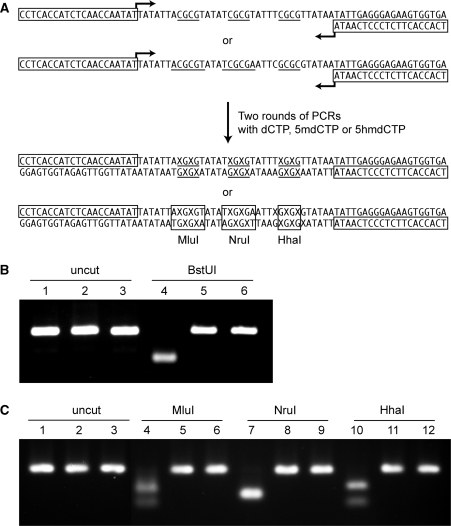
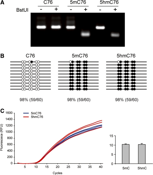
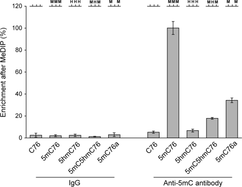
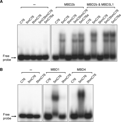
DNA cytosine-5 methylation is a well-studied epigenetic pathway implicated in gene expression control and disease pathogenesis. Different technologies have been developed to examine the distribution of 5-methylcytosine (5mC) in specific sequences of the genome. Recently, substantial amounts of 5-hydroxymethylcytosine (5hmC), most likely derived from enzymatic oxidation of 5mC by TET1, have been detected in certain mammalian tissues. Here, we have examined the ability of several commonly used DNA methylation profiling methods to distinguish between 5mC and 5hmC. We show that techniques based on sodium bisulfite treatment of DNA are incapable of distinguishing between the two modified bases. In contrast, techniques based on immunoprecipitation with anti-5mC antibody (methylated DNA immunoprecipitation, MeDIP) or those based on proteins that bind to methylated CpG sequences (e.g. methylated-CpG island recovery assay, MIRA) do not detect 5hmC and are specific for 5mC unless both modified bases occur in the same DNA fragment. We also report that several methyl-CpG binding proteins including MBD1, MBD2 and MBD4 do not bind to sequences containing 5hmC. Selective mapping of 5hmC will require the development of unique tools for the detection of this modified base.


Figures

Figure 1.
Figure 1.
Preparation and validation of modified oligonucleotides. (A) Sequence and preparation of the 76-mers used in the assays. The synthesized DNA fragments contain three BstUI sites (5′-CGCG, underlined). Two rounds of PCR were performed to obtain C76, 5mC76 or 5hmC76 containing C, 5mC or 5hmC at CpG sites. X indicates normal C, or modified bases 5mC or 5hmC. The boxed sequences indicate the PCR primers. Oligonucleotides containing both 5mC and 5hmC were synthesized chemically. (B) Analysis of PCR products C76, 5mC76 and 5hmC76 by BstUI cleavage. PCR products were prepared and digested with the methylation-sensitive restriction enzyme BstUI, then separated and visualized by electrophoresis on 3% Nusieve GTG agarose gels. Lane 1 (C76), lane 2 (5mC76) and lane 3 (5hmC76) show clean single bands for each of the PCR products. After BstUI digestion, C76 (lane 4) was fully digested, whereas samples in lane 5 (5mC76) and lane 6 (5hmC76) resisted digestion. (C) Analysis of PCR products C76, 5mC76 and 5hmC76 by cleavage with different methylation-sensitive restriction enzymes. C76 (lanes 1, 4, 7 and 10), 5mC76 (lanes 2, 5, 8 and 11) and 5hmC76 (lanes 3, 6, 9 and 12) were left untreated (lanes 1–3) or were incubated with MluI (lanes 4–6), NruI (lanes 7–9) or HhaI (lanes 9–12).
Figure 2.
Figure 2.
Comparison of the reactivity of sodium bisulfite towards 5mC and 5hmC. (A) COBRA assay. The PCR products obtained after sodium bisulfite treatment of the C-, 5mC- and 5hmC-containing templates were analyzed by the BstUI combined bisulfite restriction analysis (COBRA) method. Bisulfite-converted 76-mers were PCR amplified and digested with the restriction enzyme BstUI (5′-CGCG), which produces digestion products only when restriction sites are not converted by bisulfite. The electrophoresis on 3% Nusieve GTG agarose gels shows that 5mC76 and 5hmC76 were clearly digested, but C76 fully resisted digestion with BstUI. (B) Bisulfite sequencing. The PCR products obtained after bisulfite conversion were cloned into the pCR2.1 TA cloning vector (Invitrogen) and ten individual clones were sequenced. The 76 base pairs of C76, 5mC76 and 5hmC76 contain six cytosines or modified cytosines (5mC or 5hmC) on each strand. The modified (unconverted) cytosines are depicted as solid black circles while the unmodified cytosines are shown as open circles. Each CpG site was counted separately. (C) Real-time PCR with templates containing 5mC and 5hmC. Blue (5mC76) and red (5hmC76) curves are for three independent reactions. In the column graph, the ct value for the two templates is shown with standard deviation.
Figure 3.
Figure 3.
DNA immunoprecipitation with anti-5mC antibody. Immunoprecipitation with an antibody against 5-methylcytidine was carried out to test the antibody’s affinity towards modified cytosines. The levels of immunocaptured 76-mers were measured using liquid scintillation counting. As a control, normal mouse IgG was used for immunoprecipitation. The oligonucleotides C76, 5mC76 and 5hmC76 were synthesized by PCR and contain C, 5mC or 5hmC at three BstUI sites as schematically indicated at the top of the Figure (see Figure 1 for sequence). Oligonucleotides 5mC5hmC76 and 5mC76a were prepared by chemical synthesis and contain 5hmC or C at the central BstUI site. Experiments were done in triplicates and the standard deviation is shown.
Figure 4.
Figure 4.
Affinity of MBD proteins towards 5mC- and 5hmC-containing oligomers. (A) Binding of MBD2b and the MBD2b/MBD3L1 complex (MIRA complex) to modified cytosines. 76-mer oligonucleotides containing symmetrically modified cytosines, 5mC or 5hmC at CpG sites were incubated with recombinant MBD2b alone (200 ng of protein) or with the MBD2b/MBD3L1 complex (100 ng of each protein). The mobility shift assay was carried out using a 5% non-denaturing polyacrylamide gel. The oligonucleotides C76, 5mC76 and 5hmC76 were synthesized by PCR and contain C, 5mC or 5hmC at three BstUI sites (see Figure 1). Oligonucleotides 5mC5hmC76 and 5mC76a were prepared by chemical synthesis and contain 5hmC or C at the central BstUI site (see Figure 3). (B) Binding of MBD1 and MBD4 to modified cytosines. These proteins bind effectively to methylated CpG sequences but do not bind to the same sequences containing 5hmC in place of 5mC.

References

    1. Holliday R, Pugh JE. DNA modification mechanisms and gene activity during development. Science. 1975;187:226–232. - PubMed
    1. Riggs AD. X inactivation, differentiation, and DNA methylation. Cytogenet. Cell. Genet. 1975;14:9–25. - PubMed
    1. Bestor TH. The DNA methyltransferases of mammals. Hum. Mol. Genet. 2000;9:2395–2402. - PubMed
    1. Esteller M. Cancer epigenomics: DNA methylomes and histone-modification maps. Nat. Rev. Genet. 2007;8:286–298. - PubMed
    1. Hahn MA, Pfeifer GP. Methods for genome-wide analysis of DNA methylation in intestinal tumors. Mutat. Res. 2009 doi:10.1016/j.mrfmm.2009.10.005. - PMC - PubMed