. 2010 Jun;38(11):e123.
doi: 10.1093/nar/gkq192. Epub 2010 Apr 5.

Recombinase mediated cassette exchange into genomic targets using an adenovirus vector

Affiliations

Recombinase mediated cassette exchange into genomic targets using an adenovirus vector

David A Sorrell et al. Nucleic Acids Res. 2010 Jun.

Abstract

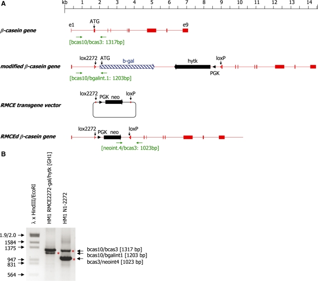
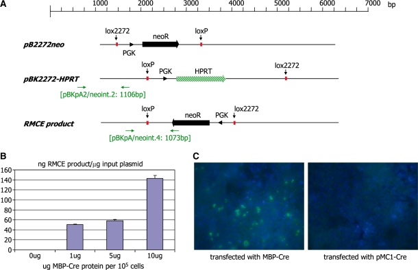
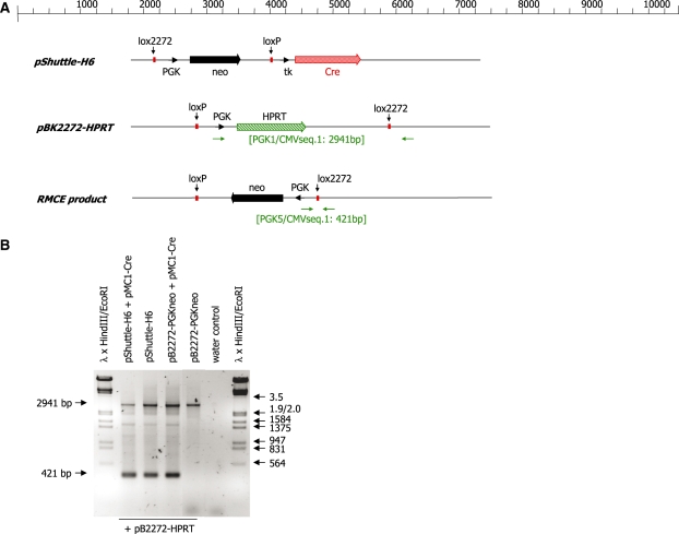
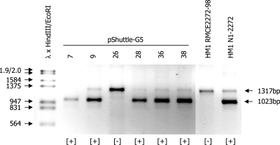
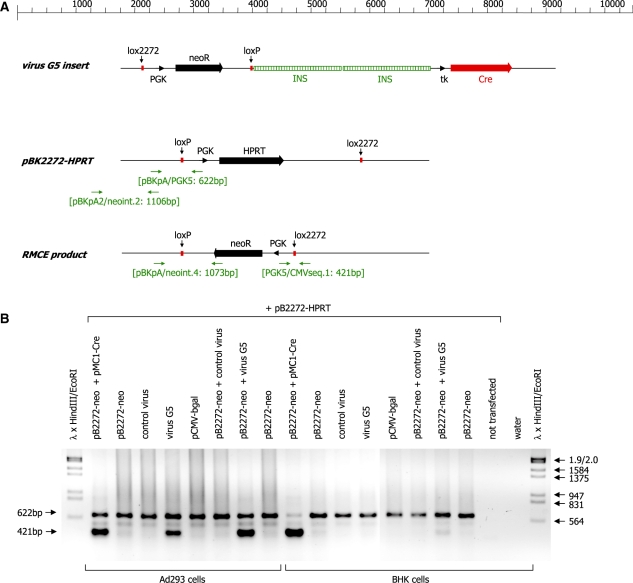
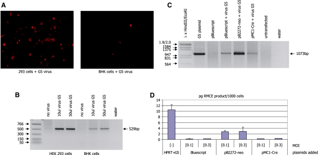
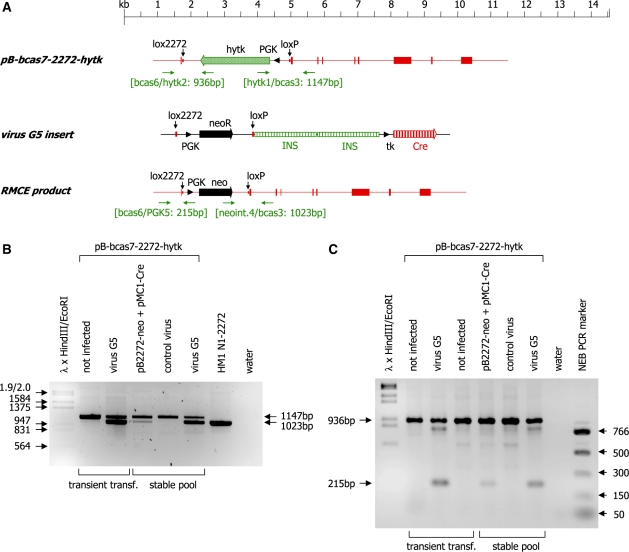
Recombinase mediated cassette exchange (RMCE) is a process in which site-specific recombinases exchange one gene cassette flanked by a pair of incompatible target sites for another cassette flanked by an identical pair of sites. Typically one cassette is present in the host genome, whereas the other gene cassette is introduced into the host cell by chemical or biological means. We show here that the frequency of cassette exchange is dependent on the relative and absolute quantities of the transgene cassette and the recombinase. We were able to successfully modify genomic targets not only by electroporation or chemically mediated gene transfer but also by using an adenovirus vector carrying both the transgene cassette to be inserted and the recombinase coding region. RMCE proceeds efficiently in cells in which the adenovirus vector is able to replicate. In contrast, insufficient quantities of the transgene cassette are produced in cells in which the virus cannot replicate. Additional transfection of the transgene cassette significantly enhances the RMCE frequency. This demonstrates that an RMCE system in the context of a viral vector allows the site directed insertion of a transgene into a defined genomic site.


Figures

Figure 1.
Figure 1.
(A) Schematic representation of the murine β-casein gene and its derivatives after homologous recombination and RMCE. Exons of the β-casein gene are indicated as solid boxes, the neomycin (neo) and hytk selection marker genes are indicated as solid arrows, respectively and the β-galactosidase gene (β-gal) is indicated as a hatched arrow. The PGK promoter elements directing expression of the selection marker genes are indicated as black arrowheads. The positions of the lox2272 and loxP sites and the translational start codon (ATG) are marked by vertical arrows. The primer binding sites (horizontal arrows) used for genotyping and the sizes of the expected PCR products are indicated. (B) PCR analysis of genomic DNA isolated from the cell clones HM1 RMCE2272-gal/hytk (GH1), and the cell clone HM1 N1-2272 derived from it. A 1317-bp band is detected in both samples and represents the unmodified β-casein allele. HM1 RMCE2272-gal/hytk (GH1) cells carry an insertion of a β-galactosidase open reading frame and a PGK-hytk expression cassette at one of the β-casein alleles as indicated by the occurrence of a 1203-bp PCR product. Cell clone HM1 N1-2272 was derived after an RMCE event which exchanged the β-gal and hytk genes for the neo selection marker gene. The correct modification is indicated by the generation of a 1023-bp PCR product and the concomitant loss of the 1203-bp band. Phage λ DNA digested with HindIII and EcoRI was used as molecular weight marker.
Figure 2.
Figure 2.
(A) Schematic representation of the plasmids pB2272-neo and pBK2272-HPRT. The neomycin resistance marker genes (neo) are indicated as solid arrows. The PGK promoter is indicated as an arrowhead. The HPRT selection marker gene is indicated as a striped arrow. The primer binding sites (horizontal arrows) used for genotyping and the sizes of the expected PCR products are indicated. (B) Quantitative PCR analysis of recombinase mediated cassette exchange in HEK 293 cells. Cells were transfected with equimolar amounts of the plasmids pBK2272-HPRT and pB2272-neo. Twenty four hours post-plasmid transfection the cells were transfected with different amounts of an MBP-Cre fusion protein using the Proteo-Juice reagent (Novagen). DNA was isolated from cells 36 h later and analysed by real-time PCR using the primer pair neoint.2/pBKpA2 (yielding a 1106-bp fragment) to quantify the concentration of the input plasmid pBK-2272HPRT and the primer pair neoint.4/pBKpA (yielding a 1073-bp fragment) to quantify the concentration of RMCE product generated. The concentrations are presented as pg RMCE product per ng input plasmid in correlation with the amount of MBP-Cre protein transfected into 1 × 10⁵ cells. (C) Immunohistochemistry of cells transfected with the plasmids pB2272-neo and pBK2272-HPRT and the protein MBP-Cre (or the plasmid pMC1-Cre; right panel). The MBP section of the protein was detected using a 1 : 200 dilution of an MBP-specific rabbit antiserum and a goat-anti-rabbit FITC-linked secondary antiserum.
Figure 3.
Figure 3.
(A) Schematic representation of the plasmids pShuttle-H6 and pBK2272-HPRT. The neomycin resistance marker genes (neo) are indicated as solid arrows. The Cre open reading frame is indicated as a shaded arrow. The PGK and the tk promoter are indicated as arrowheads. The HPRT selection marker gene is indicated as a striped arrow. The primer binding sites (horizontal arrows) used for genotyping and the sizes of the expected PCR products are indicated. (B) PCR analysis of DNA isolated from HEK 293 cells transfected with the indicated plasmids. The 2941-bp product is generated from the non-recombined pBK2272-HPRT plasmid. The 421-bp PCR product is indicative of a recombinase mediated cassette exchange between the PGKneo cassette (derived from plasmid pB2272-neo) and the PGK-HPRT cassette. Phage λ DNA digested with HindIII and EcoRI was used as molecular weight marker.
Figure 4.
Figure 4.
PCR analysis using the primer combination bcas3, bcas10 and neoint.4 on DNA isolated from representative cell clones derived after electroporation of HM1 RMCE2272-98 cells with the plasmid pShuttle-G5 and selection of the transfected cells in medium containing 200 µg/ml of G418. The 1317-bp band is detected in all samples and represents the unmodified β-casein allele. Cell clones modified by an RMCE event, which has inserted the PGK-neo selection marker cassette, display an additional 1023-bp PCR product. Phage λ DNA digested with HindIII and EcoRI was used as molecular weight marker.
Figure 5.
Figure 5.
(A) Schematic representation of the linear plasmids pShuttle-G5, pBK2272-HPRT and the expected recombination product. The neomycin resistance marker genes (neo) are indicated as solid arrows. The Cre open reading frame is indicated as a shaded arrow. The PGK and the tk promoter are indicated as arrowheads. The copies of the β-globin insulator element (INS) are indicated as vertically striped boxes. The positions of the lox2272 and loxP sites are marked by vertical arrows. (B) PCR analysis of DNA isolated from HEK 293 and BHK cells (as indicated) transfected/infected with pBK2272-HPRT plus the indicated plasmids and virus vectors. The 622-bp PCR product is generated from the non-recombined pBK2272-HPRT plasmid. The 421-bp PCR product (derived from the primer pair PGK5/CMVseq.1) is indicative of a recombinase mediated cassette exchange between the PGK-neo cassette and the PGK-HPRT cassette. Phage λ DNA digested with HindIII and EcoRI was used as molecular weight marker.
Figure 6.
Figure 6.
(A) Red fluorescent staining in HEK 293 and BHK cells transfected with the plasmid pDSred-mito-2272-PGKneo and infected with the G5 adenovirus. Red staining of the mitochondria indicates the presence of the plasmid and the virus-derived Cre protein in the same cell. (B) PCR analysis of DNA derived from the cells in A using the primer combination DSred1R/CMVseq.1. Recombination between the two identical lox2272 sites in the plasmid, which is a prerequisite for the activation of the red fluorescent protein is indicated by the occurrence of a 529-bp band (marked by the arrow). (C) PCR analysis of DNA isolated from HEK293 cells transfected with the indicated plasmids and infected with adenovirus G5 at an MOI of 0.1. RMCE is detected using the primer pair pBKpA/neoint.4 which yields an indicative 1073 bp product (marked by the arrow). (D) Real-time PCR analysis of DNA isolated from HEK293 cells transfected with the indicated plasmids and infected with adenovirus G5 at an MOI of 0.1 or 0.3 (as indicated). RMCE is detected using the primer pair PGK5/CMVseq.1 (as in Figure 5) which yields an indicative 421-bp product. The amount of PCR product is shown as pg per 1000 cells.
Figure 7.
Figure 7.
(A) Schematic representation of the plasmid pB-bcas7-2272-hytk, the insert of the virus vector G5 and the resulting RMCE product. Exons of the β-casein gene are indicated as shaded boxes, the neomycin and hytk selection marker genes are indicated as solid and shaded arrows, respectively and the Cre ORF (Cre) is indicated as a vertically striped arrow. The PGK promoter elements directing expression of the selection marker genes are indicated as black arrowheads. The positions of the lox2272 and loxP sites are marked by vertical arrows. The primer binding sites (horizontal arrows) used for genotyping and the sizes of the expected PCR products are indicated. (B) PCR analysis of DNA isolated from HEK 293 cells transiently or stably transfected with pB-bcas7-2272-hytk. The cells were either co-transfected with the indicated plasmids or infected with the indicated virus vectors 24 h post-transfection. The 3′ end of the RMCE reaction was analysed using the primer combination bcas3/neoint.4/hytk1. Successful recombination at the loxP site is indicated by the presence of the 1023-bp PCR product. Non-recombined DNA yields a PCR product of 1147 bp. Phage λ DNA digested with HindIII and EcoRI was used as molecular weight marker. (C) The 5′ end of the RMCE reaction was analysed using the primer combination bcas6/PGK5/hytk2. Successful recombination at the lox2272 site is indicated by the presence of the 215-bp PCR product. Non-recombined DNA yields a PCR product of 936 bp. Phage λ DNA digested with HindIII and EcoRI and the NEB PCR marker (New England Biolabs) were used as molecular weight markers.

Similar articles

Cited by

References

    1. Sorrell DA, Kolb AF. Targeted modification of mammalian genomes. Biotechnol. Adv. 2005;23:431–469. - PubMed
    1. Sandrin V, Russell SJ, Cosset FL. Targeting retroviral and lentiviral vectors. Curr. Top. Microbiol. Immunol. 2003;281:137–178. - PubMed
    1. Coates CJ, Kaminski JM, Summers JB, Segal DJ, Miller AD, Kolb AF. Site-directed genome modification: derivatives of DNA-modifying enzymes as targeting tools. Trends Biotechnol. 2005;23:407–419. - PubMed
    1. Kolb AF. Genome engineering using site-specific recombinases. Cloning Stem Cells. 2002;4:65–80. - PubMed
    1. Sadelain M. Insertional oncogenesis in gene therapy: how much of a risk? Gene Ther. 2004;11:569–573. - PubMed

Publication types