Case Reports
. 2010 Jun 15;19(12):2370-9.
doi: 10.1093/hmg/ddq110. Epub 2010 Apr 6.

Mutations in MUSK causing congenital myasthenic syndrome impair MuSK-Dok-7 interaction

Affiliations
Case Reports

Mutations in MUSK causing congenital myasthenic syndrome impair MuSK-Dok-7 interaction

Ricardo A Maselli et al. Hum Mol Genet. .

Abstract

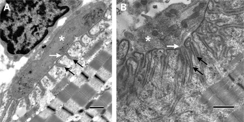
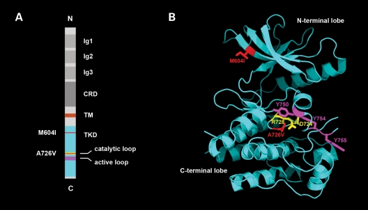
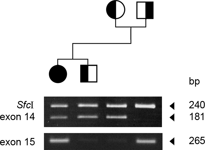
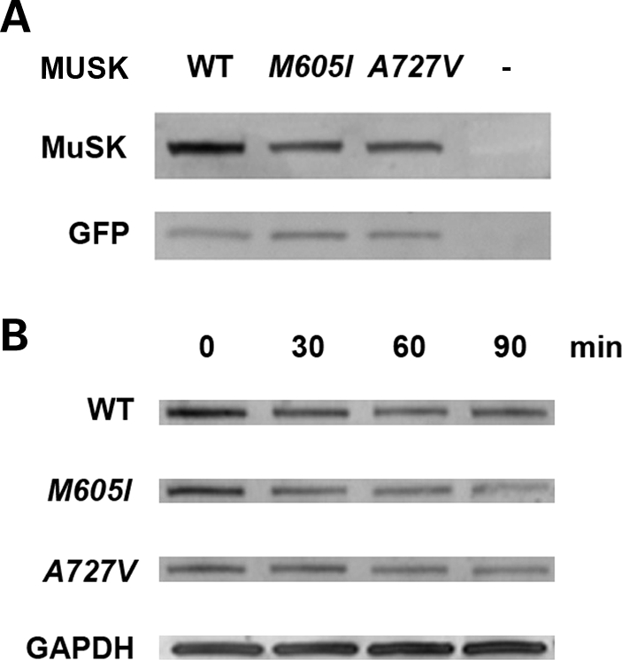
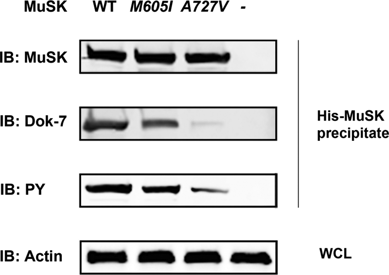
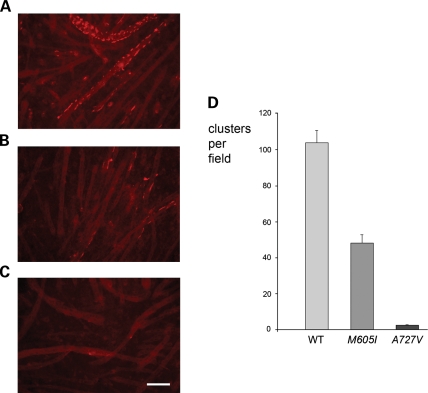
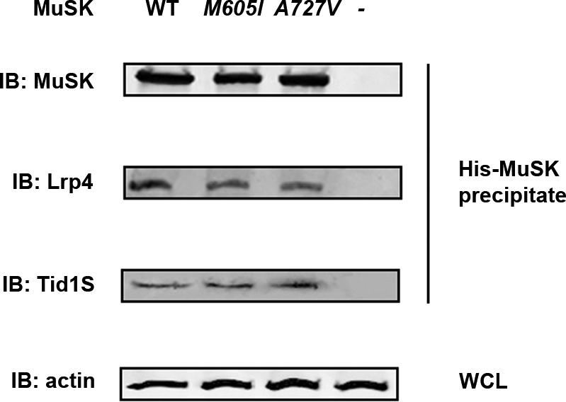
We describe a severe congenital myasthenic syndrome (CMS) caused by two missense mutations in the gene encoding the muscle specific receptor tyrosine kinase (MUSK). The identified MUSK mutations M605I and A727V are both located in the kinase domain of MuSK. Intracellular microelectrode recordings and microscopy studies of the neuromuscular junction conducted in an anconeus muscle biopsy revealed decreased miniature endplate potential amplitudes, reduced endplate size and simplification of secondary synaptic folds, which were consistent with postsynaptic deficit. The study also showed a striking reduction of the endplate potential quantal content, consistent with additional presynaptic failure. Expression studies in MuSK deficient myotubes revealed that A727V, which is located within the catalytic loop of the enzyme, caused severe impairment of agrin-dependent MuSK phosphorylation, aggregation of acetylcholine receptors (AChRs) and interaction of MuSK with Dok-7, an essential intracellular binding protein of MuSK. In contrast, M605I, resulted in only moderate impairment of agrin-dependent MuSK phosphorylation, aggregation of AChRs and interaction of MuSK with Dok-7. There was no impairment of interaction of mutants with either the low-density lipoprotein receptor-related protein, Lrp4 (a co-receptor of agrin) or with the mammalian homolog of the Drosophila tumorous imaginal discs (Tid1). Our findings demonstrate that missense mutations in MUSK can result in a severe form of CMS and indicate that the inability of MuSK mutants to interact with Dok-7, but not with Lrp4 or Tid1, is a major determinant of the pathogenesis of the CMS caused by MUSK mutations.


Figures

Figure 1.
Figure 1.
Ultrastructural findings at the NMJ. (A) An example of a NMJ from the patient demonstrating marked simplification of postsynaptic folds and underdeveloped secondary synaptic clefts (black arrows). In contrast the size of the nerve terminal (asterisk) and the width of the primary synaptic cleft (white arrow) are normal. (B) An example of a NMJ from a control showing normal secondary synaptic clefts (black arrows), nerve terminal (asterisk) and width of the primary synaptic cleft (white arrow). Calibration marks represent 1 µm.
Figure 2.
Figure 2.
Structure of MuSK showing the position of the identified mutations. (A) Schematic diagram displaying the domain organization of rat MuSK and the location of M604I (human M605I) and A726V (human A727V). The catalytic loop (amino acids 722–729), in which mutation A726V resides, is depicted in yellow. The activation loop (residues 742–763) is highlighted in purple. The positions of mutations M604I and A726V in the TKD are highlighted in red. Ig1-3, immunoglobulin-like domains; CRD, cysteine-rich domain; TM, transmembrane helix. (B) Crystal structure of the MuSK cytoplasmic domain (PDB code 1LUF, 21). M604I, replacement of methionine by isoleucine, is located in the third beta-strand of the N-terminal lobe of the TKD. The isoleucine is colored red (M604I). A726V replacement of alanine by valine is located in the catalytic loop in the C-terminal lobe of the TKD. The valine is colored red. Asp-724 and Arg-728, which are essential catalytic residues (catalytic loop), are colored yellow. Tyr-750, Tyr-754 and Tyr-755 (colored magenta) are autophosphorylation sites in the activation loop.
Figure 3.
Figure 3.
Pedigree of the family and the results of the restriction digest and allele-specific PCR. Because M605I results in a gain of a restriction site, digestion of exon 14 with SfcI results in an additional 181 DNA band in the patient and her unaffected brother and mother, but not in her father. In contrast, allele-specific PCR with a primer designed to amplify selectively the A727V mutant amplifies only the DNA from the patient and her father.
Figure 4.
Figure 4.
Expression and stability of MuSK mutants in HEK 293 cells. (A) A western blot analysis in cells co-transfected with WT or MUSK mutants and GFP using a polyclonal anti-MuSK goat antibody demonstrates reduced expression of MuSK in cells transfected with M605I and A727V in comparison with cells transfected with the WT construct (82% of WT for M605I and 87% of WT for A727V). The expression of GFP, tested with an anti-GFP polyclonal rabbit antibody, was similar in cells transfected with WT and MUSK mutants. (B) Analysis conducted in cells exposed to cycloheximide at set times intervals: 0, 30, 60 and 90 min, 36 h after transfection with WT or MUSK mutants revealed decreased expression of MuSK in cells transfected with the mutants compared with cells transfected with the WT construct. At time 0, expression of M605I was 87%, whereas A727V was 63% compared with WT. However, there were no major differences in the rate of degradation of MuSK in cells transfected with WT or MUSK mutant constructs. Expression of GAPDH detected with an anti-GAPDH monoclonal antibody showed no reduction of GAPDH expression in all the samples.
Figure 5.
Figure 5.
Interaction of MuSK with Dok-7 and MuSK phosphorylation in MuSK−/− myotubes exposed to agrin. The MuSK protein complex was eluted from lysates of MuSK−/− myotubes co-transfected with mouse plasmids expressing either His-WT-MuSK or MuSK mutants using a His SpinTrap affinity column and was subjected to immunoblot (IB) analysis using antibodies against MuSK, Dok-7 and phosphotyrosine (PY). The cells transfected with WT-MuSK showed strong Dok-7 and PY bands indicating that Dok-7 directly interacts with MuSK and induced MuSK tyrosine phosphorylation. In contrast, cells transfected with MuSK-A727V showed very weak Dok-7 and PY bands indicating failure of Dok-7 to interact with the MuSK mutant and to induce phosphorylation of the MuSK protein. The mutant MuSK-M605I showed a moderate interaction with Dok-7 and modest MuSK phosphorylation. The WCL was incubated with a monoclonal antibody directed against β-actin as a positive protein expression control.
Figure 6.
Figure 6.
Agrin-induced clustering of AChRs in MuSK−/− myotubes transfected with mouse WT- or mutant-MuSK. (A) MuSK−/− myotubes transfected with WT-MuSK showed robust clustering of AChRs. (B) MuSK−/− myotubes transfected with MuSK-M605I showed reduced clustering of AChRs. (C) MuSK−/− myotubes transfected with MuSK-A727V showed almost no clustering of AChRs. (D) Bar-graph showing the number of clusters per field in MuSK−/− myotubes transfected with mouse WT- and mutant-MuSK in four experiments involving four wells per experimental condition and the analysis of 12 fields per group. Similar protein expression for all experimental conditions was verified by Western blot analysis using an antibody against MuSK (Supplementary Material, Fig. S2). Although the difference in clustering between MuSK−/− myotubes transfected with MuSK-M605I and those transfected with WT-MuSK was significant at the P < 0.05 level (Student t-test), the difference in clustering between MuSK−/− myotubes transfected with MuSK-A727V and those transfected with WT-MuSK was significant at the P < 0.001 level (Student t-test). Calibration mark represents 100 µm
Figure 7.
Figure 7.
Interaction of mouse MuSK with Lrp4 and Tid1. The His-tagged MuSK protein complex eluted from MuSK−/− cells transfected with His-WT-MUSK or His-MuSK mutants (M605I or A727V) were subject to IB analysis using antibodies against MuSK, Lrp4 and Tid1. The WCL was used to detect β-actin as a loading control. The immunoreactivity to Lrp4 or Tid1 was not different in cells transfected with WT-MuSK or either one of the MuSK mutants. The WCL incubated with a monoclonal antibody against β-actin was consistent with equal amounts of loaded protein.

Similar articles

Cited by

References

    1. Hantaï D., Richard P., Koenig J., Eymard B. Congenital myasthenic syndromes. Curr. Opin. Neurol. 2004;17:539–551. doi:10.1097/00019052-200410000-00004. - DOI - PubMed
    1. Engel A.G., Shen X.M., Selcen D., Sine S.M. What have we learned from the congenital myasthenic syndromes. J. Mol. Neurosci. 2010;40:143–153. - PMC - PubMed
    1. Beeson D., Webster R., Cossins J., Lashley D., Spearman H., Maxwell S., Slater C.R., Newsom-Davis J., Palace J., Vincent A. Congenital myasthenic syndromes and the formation of the neuromuscular junction. Ann. N. Y. Acad. Sci. 2008;1132:99–103. doi:10.1196/annals.1405.049. - DOI - PubMed
    1. Engel A.G., Shen X.M., Selcen D., Sine S.M. Further observations in congenital myasthenic syndromes. Ann. N. Y. Acad. Sci. 2008;1132:104–113. doi:10.1196/annals.1405.039. - DOI - PMC - PubMed
    1. Chevessier F., Faraut B., Ravel-Chapuis A., Richard P., Gaudon K., Bauché S., Prioleau C., Herbst R., Goillot E., Ioos C., et al. MUSK, a new target for mutations causing congenital myasthenic syndrome. Hum. Mol. Genet. 2004;13:3229–3240. doi:10.1093/hmg/ddh333. - DOI - PubMed

Publication types

MeSH terms