. 2010 Jun 4;285(23):17359-70.
doi: 10.1074/jbc.M109.097188. Epub 2010 Apr 6.

Identification and functional characterization of a novel mitochondrial carrier for citrate and oxoglutarate in Saccharomyces cerevisiae

Affiliations

Identification and functional characterization of a novel mitochondrial carrier for citrate and oxoglutarate in Saccharomyces cerevisiae

Alessandra Castegna et al. J Biol Chem. .

Abstract

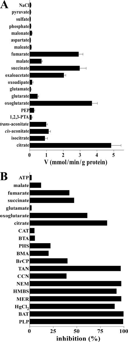
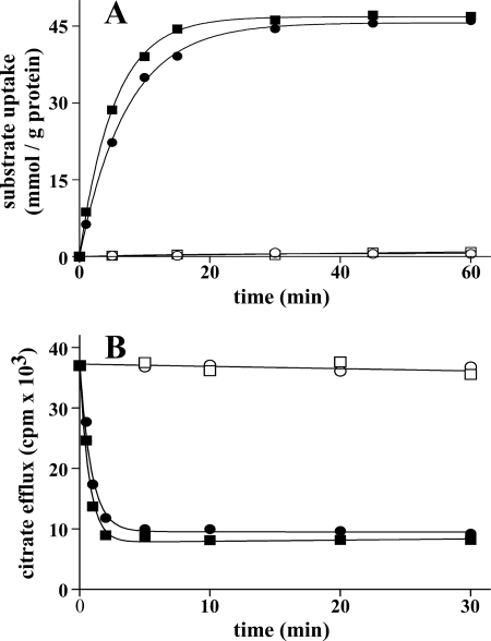
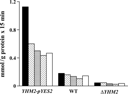
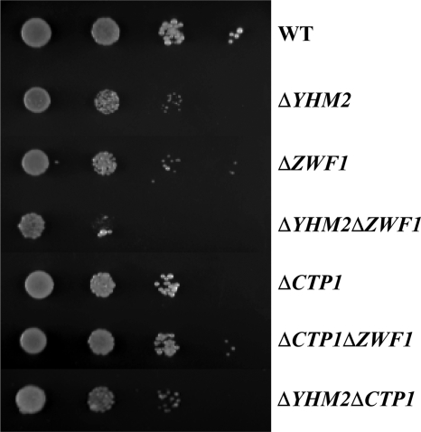
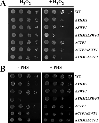
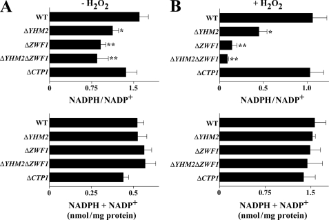
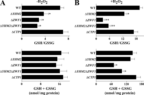
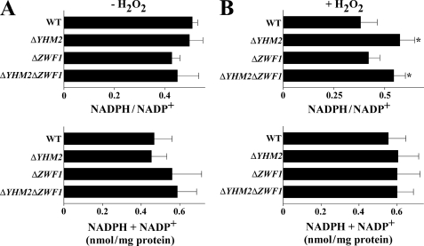
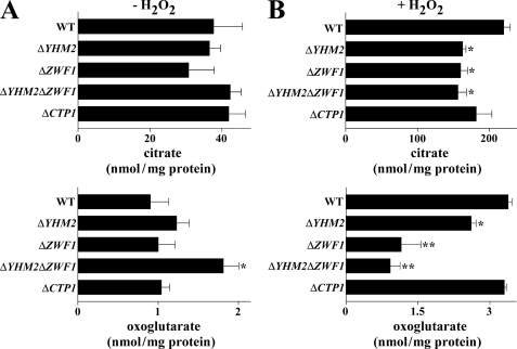
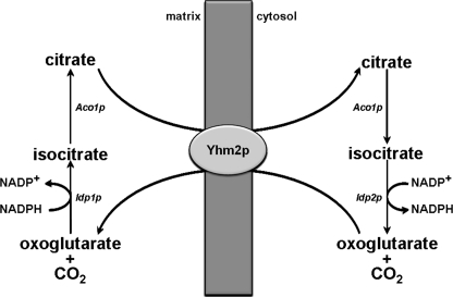
Mitochondrial carriers are a family of transport proteins that shuttle metabolites, nucleotides, and coenzymes across the mitochondrial membrane. The function of only a few of the 35 Saccharomyces cerevisiae mitochondrial carriers still remains to be uncovered. In this study, we have functionally defined and characterized the S. cerevisiae mitochondrial carrier Yhm2p. The YHM2 gene was overexpressed in S. cerevisiae, and its product was purified and reconstituted into liposomes. Its transport properties, kinetic parameters, and targeting to mitochondria show that Yhm2p is a mitochondrial transporter for citrate and oxoglutarate. Reconstituted Yhm2p also transported oxaloacetate, succinate, and fumarate to a lesser extent, but virtually not malate and isocitrate. Yhm2p catalyzed only a counter-exchange transport that was saturable and inhibited by sulfhydryl-blocking reagents but not by 1,2,3-benzenetricarboxylate (a powerful inhibitor of the citrate/malate carrier). The physiological role of Yhm2p is to increase the NADPH reducing power in the cytosol (required for biosynthetic and antioxidant reactions) and probably to act as a key component of the citrate-oxoglutarate NADPH redox shuttle between mitochondria and cytosol. This protein function is based on observations documenting a decrease in the NADPH/NADP(+) and GSH/GSSG ratios in the cytosol of DeltaYHM2 cells as well as an increase in the NADPH/NADP(+) ratio in their mitochondria compared with wild-type cells. Our proposal is also supported by the growth defect displayed by the DeltaYHM2 strain and more so by the DeltaYHM2DeltaZWF1 strain upon H(2)O(2) exposure, implying that Yhm2p has an antioxidant function.


Figures

FIGURE 1.
FIGURE 1.
Substrate specificity of recombinant and reconstituted Yhm2p. A, dependence of Yhm2p activity on internal substrate. Proteoliposomes were preloaded internally with various substrates (concentration, 20 mm). Transport was initiated by adding 0.15 mm [14C]citrate to proteoliposomes and terminated after 1 min. Values are means ± S.E. of at least three independent experiments. Abbreviations used are as follows: 1,2,3-PTA, 1,2,3-pentanetricarboxylate; PEP, phosphoenolpyruvate. B, effect of inhibitors and external substrates on the [14C]citrate/citrate exchange catalyzed by reconstituted Yhm2p. Proteoliposomes were preloaded internally with 20 mm citrate, and transport was initiated by adding 0.15 mm [14C]citrate. The incubation time was 1 min. Thiol reagents and α-cyanocinnamate were added 2 min before the labeled substrate; the other indicated inhibitors and substrates were added together with [14C]citrate. The final concentrations of the inhibitors were 0.1 mm (HMBS, p-hydroxymercuribenzene sulfonate; HgCl2, mercuric chloride; MER, mersalyl), 10 mm (PLP, pyridoxal 5′-phosphate; BAT, bathophenanthroline), 1 mm (NEM, N-ethylmaleimide; CCN, α-cyanocinnamate), 2 mm (BTA, benzene 1,2,3-tricarboxylate; BMA, butylmalonate; PHS, phenylsuccinate), 0.2 mm (BrCP, bromcresol purple), 0.05% (TAN, tannic acid), and 10 μm (CAT, carboxyatractyloside). The final concentration of unlabeled substrates added together with [14C]citrate was 3 mm. The extents of inhibition (%) from a representative experiment are given. Similar results were obtained in at least three independent experiments.
FIGURE 2.
FIGURE 2.
Kinetics of [14C]citrate and [14C]oxoglutarate transport in proteoliposomes reconstituted with Yhm2p. A, uptake of citrate or oxoglutarate. 2 mm [14C]citrate or [14C]oxoglutarate was added to proteoliposomes containing 20 mm citrate (■) or oxoglutarate (●), respectively (exchange), or 10 mm NaCl and no substrate for either citrate (□) or oxoglutarate (○) uptake (uniport). B, efflux of [14C]citrate from proteoliposomes reconstituted in the presence of 1 mm citrate. The internal substrate pool was labeled with [14C]citrate by carrier-mediated exchange equilibration. Then the proteoliposomes were passed through Sephadex G-75. The efflux of [14C]citrate was started by adding buffer G alone (□), 5 mm citrate (■), or oxoglutarate (●) in buffer G or 5 mm citrate, 30 mm pyridoxal 5′-phosphate, and 10 mm bathophenanthroline in buffer G (○). Similar results were obtained in three independent experiments for both uptake and efflux.
FIGURE 3.
FIGURE 3.
Citrate exchange activities in liposomes reconstituted with mitochondrial extracts from the ΔYHM2 strain, parental strain, and the parental strain transformed with the YHM2-pYES2 plasmid. These strains were grown as indicated under “Experimental Procedures” for YHM2-pYES2 cells. Mitochondria were isolated and solubilized with 1% Triton X-100, 50 mm NaCl, and 10 mm PIPES, pH 7.0. The extracts (30 μg of protein) were reconstituted into liposomes preloaded with 20 mm citrate (black columns), oxoglutarate (hatched columns), succinate (gray columns), oxaloacetate (inverted hatched columns), or fumarate (white columns). Transport was started by adding 0.15 mm [14C]citrate and terminated after 15 min. WT, wild type.
FIGURE 4.
FIGURE 4.
Growth behavior of ΔYHM2, ΔZWF1, ΔYHM2ΔZWF1, ΔCTP1, ΔCTP1ΔZWF1, and ΔYHM2ΔCTP1 strains in acetate-supplemented SM. 4-Fold serial dilutions of equally numbered wild-type (WT), ΔYHM2, ΔZWF1, ΔYHM2ΔZWF1, ΔCTP1, ΔCTP1ΔZWF1, and ΔYHM2ΔCTP1 cells (precultured overnight in YP with glucose) were plated on solid acetate-supplemented SM at 30 °C.
FIGURE 5.
FIGURE 5.
Growth behavior of ΔYHM2, ΔZWF1, ΔYHM2ΔZWF1, ΔCTP1, ΔCTP1ΔZWF1, and ΔYHM2ΔCTP1 strains in acetate-supplemented SC in the presence or absence of H2O2 (A) and phytosphingosine (B). 4-Fold serial dilutions of equally numbered wild-type (WT), ΔYHM2, ΔZWF1, ΔYHM2ΔZWF1, ΔCTP1, ΔCTP1ΔZWF1, and ΔYHM2ΔCTP1 cells (precultured overnight in YP with glucose) were plated on solid acetate-supplemented SC with or without 1.25 mm H2O2 at 30 °C (A) and 5 μm phytosphingosine (PHS) at 37 °C (B).
FIGURE 6.
FIGURE 6.
NADPH/NADP+ ratios in the cytosol of parental and deleted S. cerevisiae strains. Yeast strains, precultured in YP with glucose and grown in acetate-supplemented SC, were incubated in the presence (B) or absence (A) of H2O2 for 60 min. NADPH/NADP+ ratios and the sums of NADPH + NADP+ are reported as means ± S.E. of at least three independent experiments. Differences between the NADPH/NADP+ ratio of ΔYHM2, ΔZWF1, and ΔYHM2ΔZWF1 cells and control (wild-type (WT) cells), with and without H2O2, were significant (*, p < 0.05, and **, p < 0.01, one-way ANOVA followed by Bonferroni's t test).
FIGURE 7.
FIGURE 7.
GSH/GSSG ratios in the cytosol of parental and deleted S. cerevisiae strains. Yeast strains, precultured in YP with glucose and grown in acetate-supplemented SC, were incubated in the presence (B) or absence (A) of H2O2 for 60 min. GSH/GSSG ratios and the sums of GSH + GSSG are reported as means ± S.E. of at least three independent experiments. Differences between the GSH/GSSG ratio of ΔYHM2, ΔZWF1, and ΔYHM2ΔZWF1 cells and control (wild-type (WT) cells), with and without H2O2, were significant (*, p < 0.05, and **, p < 0.01, one-way ANOVA followed by Bonferroni's t test). Differences between the sum of GSH + GSSG of ΔZWF1 and ΔYHM2ΔZWF1 and control (wild-type cells) in the presence of H2O2 were significant (*, p < 0.05, one-way ANOVA followed by Bonferroni's t test).
FIGURE 8.
FIGURE 8.
NADPH/NADP+ ratios in the mitochondria of parental and deleted S. cerevisiae strains. Yeast strains, precultured in YP with glucose and grown in acetate-supplemented SC, were incubated in the presence (B) or absence (A) of H2O2 for 60 min. NADPH/NADP+ ratios and the sums of NADPH + NADP+ are reported as means ± S.E. of at least three independent experiments. Differences between the NADPH/NADP+ ratio of ΔYHM2 and ΔYHM2ΔZWF1 cells and control (wild-type (WT) cells) in the presence of H2O2 were significant (*, p < 0.05, one-way ANOVA followed by Bonferroni's t test).
FIGURE 9.
FIGURE 9.
Citrate and oxoglutarate levels in the cytosol of parental and deleted S. cerevisiae strains. Yeast strains, precultured in YP with glucose and grown in acetate-supplemented SC, were incubated in the presence (B) or absence (A) of H2O2 for 60 min. Means ± S.E. of at least three independent experiments are reported. Differences between cytosolic citrate of ΔYHM2, ΔZWF1, and ΔYHM2ΔZWF1 cells and control (wild-type (WT) cells) in the presence of H2O2 were significant (*, p < 0.05, one-way ANOVA followed by Bonferroni's t test). The difference between cytosolic oxoglutarate of ΔYHM2ΔZWF1 and wild-type cells in the absence of H2O2 is significant (*, p < 0.05). Differences between cytosolic oxoglutarate of ΔYHM2, ΔZWF1, and ΔYHM2ΔZWF1 cells and wild-type cells in the presence of H2O2 were significant (*, p < 0.05, and **, p < 0.01, one-way ANOVA followed by Bonferroni's t test).
FIGURE 10.
FIGURE 10.
Cell antioxidant response to oxidative stress. DCFH-DA-preloaded wild-type (■), ΔYHM2 (○), ΔZWF1 (□), and ΔYHM2ΔZWF1 (▵) cells were incubated in acetate-supplemented SC in the presence of H2O2 for the indicated time periods. DCF fluorescence was measured using a microplate reader and expressed as the percentage of the corresponding wild-type values. Values are means ± S.E. of four independent experiments.
FIGURE 11.
FIGURE 11.
Flow cytometric analysis of ROS levels in wild-type and knock-out S. cerevisiae strains. DCFH-DA-preloaded wild-type (WT), ΔYHM2, ΔZWF1, and ΔYHM2ΔZWF1 cells were incubated in acetate-supplemented SC in the presence or absence of H2O2 for the time periods indicated. Results from a representative experiment are shown as cell number (counts) versus DCF fluorescence.
FIGURE 12.
FIGURE 12.
Scheme of the reactions involved in the citrate-oxoglutarate NADPH shuttle from the mitochondrial matrix to the cytosol in S. cerevisiae.

Similar articles

Cited by

References

    1. Cho J. H., Ha S. J., Kao L. R., Megraw T. L., Chae C. B. (1998) Mol. Cell. Biol. 18, 5712–5723 - PMC - PubMed
    1. MacAlpine D. M., Perlman P. S., Butow R. A. (1998) Proc. Natl. Acad. Sci. U.S.A. 95, 6739–6743 - PMC - PubMed
    1. Palmieri F. (2004) Pflugers Arch. 447, 689–709 - PubMed
    1. Palmieri F. (2008) Biochim. Biophys. Acta 1777, 564–578 - PubMed
    1. Palmieri F., Pierri C. L. (2010) FEBS Lett. 584, 1931–1939 - PubMed

Publication types

MeSH terms