. 2010 Jun 4;285(23):17869-79.
doi: 10.1074/jbc.M110.101055. Epub 2010 Apr 6.

MicroRNA-155 regulates cell survival, growth, and chemosensitivity by targeting FOXO3a in breast cancer

Affiliations

MicroRNA-155 regulates cell survival, growth, and chemosensitivity by targeting FOXO3a in breast cancer

William Kong et al. J Biol Chem. .

Retraction in

Abstract

Breast cancer is the second leading cause of cancer death in women. Despite improvement in treatment over the past few decades, there is an urgent need for development of targeted therapies. miR-155 (microRNA-155) is frequently up-regulated in breast cancer. In this study, we demonstrate the critical role of miR-155 in regulation of cell survival and chemosensitivity through down-regulation of FOXO3a in breast cancer. Ectopic expression of miR-155 induces cell survival and chemoresistance to multiple agents, whereas knockdown of miR-155 renders cells to apoptosis and enhances chemosensitivity. Further, we identified FOXO3a as a direct target of miR-155. Sustained overexpression of miR-155 resulted in repression of FOXO3a protein without changing mRNA levels, and knockdown of miR-155 increases FOXO3a. Introduction of FOXO3a cDNA lacking the 3'-untranslated region abrogates miR-155-induced cell survival and chemoresistance. Finally, inverse correlation between miR-155 and FOXO3a levels were observed in a panel of breast cancer cell lines and tumors. In conclusion, our study reveals a molecular link between miR-155 and FOXO3a and presents evidence that miR-155 is a critical therapeutic target in breast cancer.


Figures
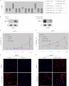
FIGURE 1.
FIGURE 1.
miR-155 induces breast cancer cell growth and survival and chemoresistance. A, miR-155 and U6 expression in 12 breast cancer cell lines was determined by real-time PCR. Relative expression was calculated by obtaining ΔCT, where the CT value of miR-155 was divided by the U6 CT from the same sample. The resulting value was then transformed by applying 2−(ΔCT). B and C, Northern blot. BT-474, expressing low endogenous miR-155, was stably infected with lentivirus expressing miR-155 and vector (B). HS578T cells, presenting high endogenous miR-155, were transfected with anti-miR-155 (ASO) and control oligonucleotide (C). After incubation of 24 h, total RNAs were isolated and subjected to Northern blot analysis with the indicated probes. D–F, expression of miR-155 induces cell growth, survival, and chemoresistance. miR-155- and vector-infected or oligonucleotide mimic-transfected BT-474 cells were treated with and without doxorubicin for the indicated times (D) or with the indicated chemotherapeutic agents for 24 h (E and F). The growth curve was determined by counting cell number at different time points (D). Apoptosis was assayed with a terminal deoxynucleotidyltransferase-mediated dUTP nick end labeling assay and Cell Death Detection ELISAPLUS kit (E) as well as caspase 3/7 activity (F). G–I, knockdown of miR-155 inhibits cell growth/survival and sensitizes cells to chemotherapeutic drug-induced cell growth arrest and apoptosis. Anti-miR-155 and scrambled oligonucleotide-treated HS578T cells were treated with the same agents and assayed with the same methods as described in D–F. Data represent three independent experiments with four replicates each. *, p < 0.05. Error bars, S.E.
FIGURE 1.
FIGURE 1.
miR-155 induces breast cancer cell growth and survival and chemoresistance. A, miR-155 and U6 expression in 12 breast cancer cell lines was determined by real-time PCR. Relative expression was calculated by obtaining ΔCT, where the CT value of miR-155 was divided by the U6 CT from the same sample. The resulting value was then transformed by applying 2−(ΔCT). B and C, Northern blot. BT-474, expressing low endogenous miR-155, was stably infected with lentivirus expressing miR-155 and vector (B). HS578T cells, presenting high endogenous miR-155, were transfected with anti-miR-155 (ASO) and control oligonucleotide (C). After incubation of 24 h, total RNAs were isolated and subjected to Northern blot analysis with the indicated probes. D–F, expression of miR-155 induces cell growth, survival, and chemoresistance. miR-155- and vector-infected or oligonucleotide mimic-transfected BT-474 cells were treated with and without doxorubicin for the indicated times (D) or with the indicated chemotherapeutic agents for 24 h (E and F). The growth curve was determined by counting cell number at different time points (D). Apoptosis was assayed with a terminal deoxynucleotidyltransferase-mediated dUTP nick end labeling assay and Cell Death Detection ELISAPLUS kit (E) as well as caspase 3/7 activity (F). G–I, knockdown of miR-155 inhibits cell growth/survival and sensitizes cells to chemotherapeutic drug-induced cell growth arrest and apoptosis. Anti-miR-155 and scrambled oligonucleotide-treated HS578T cells were treated with the same agents and assayed with the same methods as described in D–F. Data represent three independent experiments with four replicates each. *, p < 0.05. Error bars, S.E.
FIGURE 2.
FIGURE 2.
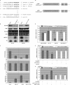
miR-155 targets FOXO3a through interaction with FOXO3a 3′-UTR. A, sequence alignment of the human miR-155 sequences with a region of the FOXO3a 3′-UTR from indicated species. Conserved nucleotides are in boldface type (top). The bottom panel shows a mutant of FOXO3a 3′-UTR for pMIR-REPORT, and the mutant nucleotides are underlined. B, expression of miR-155 reduces FOXO3a protein but not mRNA levels. BT-474 cells were transfected with pre-miR-155 or control oligonucleotide and then subjected to Western blot (top two panels), RT-PCR (panels 3 and 4), and Northern blot (panels 5 and 6) analyses. The bottom panels show quantifications of levels of FOXO3a and miR-155. C, knockdown of miR-155 induces FOXO3a protein level. Hs578T cells were transfected with miR-155 ASO and control oligonucleotide for 24 h and then examined for FOXO3a and miR-155 levels as described in B. Actin was used as loading control for Western and RT-PCR analysis. miR-155 was normalized by U6. D, tandem repeats of depicted sequences of WT and MUT FOXO3a 3′-UTR were used for cloning into pMIR-REPORT vector downstream of the luciferase gene. The cloning strategy is detailed under “Experimental Procedures.” E–G, luciferase reporter assay. The indicated cells were transfected with the indicated plasmids and oligonucleotide as well as β-galactosidase. After a 24-h incubation, cells were subjected to a luciferase assay. Luciferase activity was normalized with β-galactosidase. Relative luciferase activities are presented. Data represent three independent experiments in triplicate. *, p < 0.05. Error bars, S.E.
FIGURE 3.
FIGURE 3.
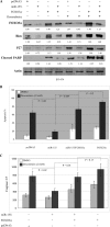
miR-155 inhibits FOXO3a downstream targets, Bim and p27, and modulates doxorubicin effects on FOXO3a/Bim/p27 and apoptosis. Lenti-miR-155-infected and vector-stably infected BT-474 cells were treated with doxorubicin at the indicated time points and then subjected to immunoblotting analysis with the indicated antibodies (A) and apoptosis assay (B). C and D, control and anti-miR-155 oligonucleotide-treated HS578T cells were treated and analyzed as described in A and B. Apoptosis experiments were repeated three times in triplicate. Error bars, S.E.
FIGURE 4.
FIGURE 4.
Transfection of FOXO3a cDNA lacking 3′-UTR overrides miR-155 effects on Bim and p27 expression and cell survival. BT-474 cells were transfected/treated with and without indicated constructs and agent and then immunoblotted with the indicated antibodies and quantified as described under “Experimental Procedures” (A). Further, following treatment with and without doxorubicin, cells were also assayed for apoptosis (B) and caspase 3/7 activity (C). *, p < 0.05. Error bars, S.E.
FIGURE 5.
FIGURE 5.
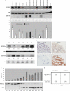
miR-155 inversely correlates with FOXO3a expression in breast cancer tissue and cell lines. A, Western (top two panels) and Northern (panels 3 and 4) analyses of 12 breast cell lines for expression of FOXO3a and miR-155. The bottom panel shows the quantification of FOXO3a and miR-155. B and C, expression of FOXO3a and miR-155 was analyzed in representative of breast tumors with Western/Northern blot (B), miR-155 LNA-ISH, and immunohistochemical staining (C). D, representatives of recurrent tumors (RT1–RT10) were analyzed with qRT-PCR (top; miR-155) and immunoblotting analysis (bottom). Expression levels of miR-155 and FOXO3a were quantified as described under “Experimental Procedures.” E, χ2 test analysis of miR-155 and FOXO3a expression in 77 breast cancer specimens was examined. The inverse correlation is significant (p < 0.001). Error bars, S.E.

Similar articles

Cited by

References

    1. Bartel D. P. (2009) Cell 136, 215–233 - PMC - PubMed
    1. Lagos-Quintana M., Rauhut R., Lendeckel W., Tuschl T. (2001) Science 294, 853–858 - PubMed
    1. Lau N. C., Lim L. P., Weinstein E. G., Bartel D. P. (2001) Science 294, 858–862 - PubMed
    1. Lee R. C., Feinbaum R. L., Ambros V. (1993) Cell 75, 843–854 - PubMed
    1. Calin G. A., Dumitru C. D., Shimizu M., Bichi R., Zupo S., Noch E., Aldler H., Rattan S., Keating M., Rai K., Rassenti L., Kipps T., Negrini M., Bullrich F., Croce C. M. (2002) Proc. Natl. Acad. Sci. U.S.A. 99, 15524–15529 - PMC - PubMed

Publication types