. 2010 Jun 4;285(23):17789-97.
doi: 10.1074/jbc.M109.082057. Epub 2010 Apr 6.

Identification of heterogeneous nuclear ribonucleoprotein K as a transactivator for human low density lipoprotein receptor gene transcription

Affiliations

Identification of heterogeneous nuclear ribonucleoprotein K as a transactivator for human low density lipoprotein receptor gene transcription

Hai Li et al. J Biol Chem. .

Abstract

hnRNP K, a member of the family of heterogeneous ribonucleoproteins, is known to exert various functional roles in the nucleus, cytoplasm, and mitochondria to affect different cellular processes including chromatin remodeling, transcription, splicing, and translation. Here we report, for the first time, that hnRNP K is specifically involved in human LDL receptor (LDLR) gene transcription in HepG2 cells. We show that depletion of hnRNP K by siRNA transfection reduces the expression of LDLR mRNA and protein by more than 50% as measured by quantitative real-time PCR and Western blot analysis. Importantly, we show that the decay rate of LDLR mRNA is not affected by hnRNP K siRNA transfection, whereas the LDLR promoter activity is significantly decreased. Furthermore, overexpression of hnRNP K increased the LDLR promoter activity by the luciferase reporter assay. By utilizing a series of mutational and deletional constructs of LDLR promoter luciferase reporters, we mapped the K-responsive element to the repeat 3 (R3) sequence of the LDLR promoter. Electrophoretic mobility shift assays show that the K protein binds to a single-stranded DNA probe containing the CT-rich element of R3, which is in contrast to the requirement of double-stranded DNA for Sp1 to bind to R3. Finally, chromatin immunoprecipitation assays reveal a direct interaction of hnRNP K with the LDLR promoter in intact HepG2 cells. These new findings provide strong evidence demonstrating that hnRNP K is an important transactivator for human LDLR gene transcription. This work sheds new light on our current understanding of how LDLR gene expression is controlled at the transcriptional level.


Figures
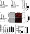
FIGURE 1.
FIGURE 1.
hnRNP K knockdown attenuates LDLR expression. A, 0.5 × 10⁶ HepG2 cells/well were plated into a 6-well cell culture plate, and 12.5 μl/well of 2 μm scrambled or hnRNP K siRNAs were simultaneously transfected into HepG2 cells for 2 days before total RNA or protein extraction. RT-PCR was performed to detect hnRNP K and GAPDH mRNA, respectively. B, total protein was extracted from HepG2 cells and hnRNP K and β-actin were detected by Western blotting, respectively. C, real-time PCR was performed to measure the LDLR mRNA levels from cDNA samples obtained from A. LDLR mRNA levels were normalized by GAPDH mRNA expression levels. ***, p < 0.001. The data shown are average of three separate transfections. D, Western blotting was performed to detect the LDLR and β-actin protein levels in both scrambled or hnRNP K siRNA-transfected samples. E, HepG2 cells were seeded onto 6-well plates and transfected with siRNAs for 48 h; receptor-mediated uptake of DiI-LDL was examined with a fluorescent microscope after incubation with DiI-LDL (5 μg/ml) for 4 h. F, the uptake of DiI-LDL uptake in HepG2 cells was further analyzed by FACS. The mean fluorescence value (MFV) of control cells transfected with scrambled siRNA was defined as 100%, and the MFV in hnRNP K siRNA-transfected cells were normalized by that value. ***, p < 0.001. The data shown are the average of two independent experiments. G, real-time PCR analysis of mRNA expression of K target genes and nontargeted genes using cDNA samples obtained from A. ***, p < 0.001. H, cell proliferation rates were determined using the CellTiter-Glo Luminescent Cell Viability Assay kit as described under “Experimental Procedures.”
FIGURE 2.
FIGURE 2.
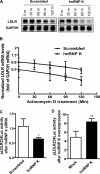
Transcriptional regulation of LDLR expression by hnRNP K. A, HepG2 cells were seeded onto 6-well cell culture plates, and total RNA was extracted as described in the legend to Fig. 1 except that actinomycin D (5 μg/ml) was added and incubated for the indicated periods of time before collecting cells. 10 μg/sample of total RNA was loaded onto a 1.2% denatured agarose gel. LDLR and GAPDH transcripts were detected by 32P-labeled DNA probes, respectively. B, the density of LDLR and GAPDH mRNA bands was calculated and LDLR mRNA levels were normalized by corresponding GAPDH values. Linear degradation curves were obtained by plotting the relative LDLR mRNA values against time. The data represent the average of two independent experiments. C, HepG2 cells were transfected by scrambled or hnRNP K siRNA on the day when cells were seeded. pLDLR234Luc and pRL-TK were cotransfected at a ratio of 20:1 the next day and incubated for two more days before lysis. D, pLDLR234Luc together with pRL-TK at a ratio of 20:1 were transiently transfected into HepG2 cells by FuGene 6 along with pcDNA3-myc-hnRNP K or mock vector for 2 days before examining luciferase activity. Results are presented as a ratio of firefly luciferase activity to Renilla. *, p < 0.05 and **, p < 0.01 compared with control. The data shown are representative of three separate transfections with similar results.
FIGURE 3.
FIGURE 3.
The repeat 3 of the LDLR promoter is responsive to hnRNP K knockdown. A, schematic representation of the wild type, 3 mutants, and 1 deletion of pLDLR234Luc reporters. To perform siRNA knockdown and dual-luciferase assay on 96-well plates, 0.5 μl/well of 2 μm small RNA were mixed with 1.5 × 10⁴ HepG2 cells/well. The next day, 100 ng/well of pLDLR234Luc reporter plus 5 ng of pRL-TK reporter as transfection control were cotransfected into these cells. Two days after transfection with different reporters, cells were lysed. The normalized luciferase activities of various reporters are presented in B, and in C the relative luciferase activity in scrambled siRNA-transfected cells is expressed as 1. The data shown are representative of three separate transfections with similar results.
FIGURE 4.
FIGURE 4.
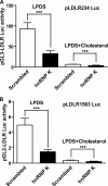
LDLR transactivation by hnRNP K is independent of intracellular cholesterol levels. A, 0.5 μl/well of 2 μm small RNA was mixed with 1.5 × 10⁴ HepG2 cells/well on 96-well cell culture plates. The next day, 100 ng/well of pLDLR234Luc reporter plus 5 ng of pRL-TK reporter as transfection control were transfected into these cells. Two days after reporters' transfection, culture medium was changed to MEM containing 10% LPDS or MEM containing 10% LPDS plus cholesterol (10 μg/ml cholesterol + 1 μg/ml 25-hydroxycholesterol), and cells were incubated for 24 h prior to cell lysis for performing luciferase activity assays. B, dual-luciferase analysis was performed as described in Fig. 4A using pLDLR1563Luc. Each value represents the mean ± S.D. of 6 wells per condition. ***, p < 0.001. The data shown are representative of three separate transfections with similar results.
FIGURE 5.
FIGURE 5.
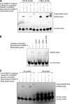
hnRNP K binds single-stranded LDLR probe containing CT-rich element of LDLR gene promoter. A, EMSA assays were carried out using 5′-biotin end-labeled single-stranded LDLR or c-myc DNA probes with LightShift® Chemiluminescent EMSA kit. Unlabeled single-stranded probes were used as specific competitors, and a DNA fragment from intron 1 of LDLR gene was used as a nonspecific competitor in 100-fold excess. To observe the specific shifted bands, recombinant hnRNP K protein was added to the reaction mixture. An antibody against hnRNP K was added into the DNA-protein reaction mixture to detect supershifted bands. B, EMSA assays were carried out to examine the ability of mutated R3D oligonucleotide to compete the K binding to the labeled probe. C, EMSA assays were carried out using 5′-biotin end-labeled single- or double-stranded LDLR promoter probes. Shifted and supershifted bands were detected by mixing recombinant hnRNP K protein or hnRNP K protein plus anti-hnRNP K antibody, respectively.
FIGURE 6.
FIGURE 6.
Sp1 only binds the double-stranded LDLR DNA probe. EMSA assays were carried out using 5′-biotin end-labeled single- or double-stranded LDLR DNA probes with recombinant purified Sp1 protein. EMSA was conducted as described in legend of Fig. 5.
FIGURE 7.
FIGURE 7.
ChIP analyses of hnRNP K and acetylated histone H3 and H4 association with the LDLR promoter in HepG2 cells. A, antibodies to hnRNP K, acetylated histone H3, H4, and HNF1β were used in a ChIP analysis followed by PCR to amplify a 180-bp region surrounding the repeat 3 sequence of LDLR promoter from genomic DNA isolated from HepG2 cells transfected with scrambled siRNA or hnRNP K siRNA. Normal rabbit IgG was included in the assay as negative controls for nonspecific binding. The data shown are representative of three independent assays with similar results. B, ChIP assay was performed to examine the binding of K to the PCSK9 promoter and exon 12 regions as nonspecific controls.

Similar articles

Cited by

References

    1. Rader D. J., Cohen J., Hobbs H. H. (2003) J. Clin. Invest. 111, 1795–1803 - PMC - PubMed
    1. Goldstein J. L., Brown M. S. (2001) Science 292, 1310–1312 - PubMed
    1. Goldstein J. L., Brown M. S. (2009) Arterioscler. Thromb. Vasc. Biol. 29, 431–438 - PMC - PubMed
    1. Smith J. R., Osborne T. F., Goldstein J. L., Brown M. S. (1990) J. Biol. Chem. 265, 2306–2310 - PubMed
    1. Liu J., Ahlborn T. E., Briggs M. R., Kraemer F. B. (2000) J. Biol. Chem. 275, 5214–5221 - PubMed

Publication types

LinkOut - more resources