. 2010 May 28;285(22):16958-66.
doi: 10.1074/jbc.M109.093005. Epub 2010 Apr 6.

Foxo3a regulates apoptosis by negatively targeting miR-21

Affiliations

Foxo3a regulates apoptosis by negatively targeting miR-21

Kun Wang et al. J Biol Chem. .

Abstract

MicroRNAs are a class of small non-coding RNAs and participate in the regulation of apoptotic program. Although miR-21 is able to inhibit apoptosis, its expression regulation and downstream targets remain to be fully elucidated. Here we report that the transcriptional factor Foxo3a initiates apoptosis by transcriptionally repressing miR-21 expression. Our results showed that doxorubicin could simultaneously induce the translocation of Foxo3a to the cell nuclei and a reduction in miR-21 expression. Knockdown of Foxo3a resulted in an elevation in miR-21 levels, whereas enforced expression of Foxo3a led to a decrease in miR-21 expression. In exploring the molecular mechanism by which Foxo3a regulates miR-21, we observed that Foxo3a bound to the promoter region of miR-21 and suppressed its promoter activity. These results indicate that Foxo3a can transcriptionally repress miR-21 expression. In searching for the downstream targets of miR-21 in apoptosis, we found that miR-21 suppressed the translation of Fas ligand (FasL), a pro-apoptotic factor. Furthermore, Foxo3a was able to up-regulate FasL expression through down-regulating miR-21. Our data suggest that Foxo3a negatively regulates miR-21 in initiating apoptosis.


Figures
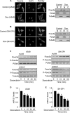
FIGURE 1.
FIGURE 1.
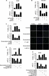
Doxorubicin induces a redistribution of Foxo3a from the cytoplasm to nuclei and a reduction of miR-21 levels. A, doxorubicin induces Foxo3a redistributions from the cytoplasm to nuclei in A549 cells. A549 cells were treated with 2 μm doxorubicin (Dox). 1 h after treatment, cells were collected for the analysis of Foxo3a by immunofluorescent staining (left panel) or by immunoblotting with the cellular fractions of nuclei (N) or cytosol (C) (right panel). Cell nuclei were stained by 4′,6-diamidino-2-phenylindole (DAPI). Bar = 10 μm. Proliferating cell nuclear antigen (PCNA) is a nucleic marker. Tubulin is a cytosolic marker. B, doxorubicin induces Foxo3a redistributions from the cytoplasm to nuclei in SH-EP1 cells. The experiments were performed as described for A except that SH-EP1 cells were used. C, doxorubicin induces a time-dependent redistribution of Foxo3a. A549 (left panel) and SH-EP1 cells (right panel) were treated with 2 μm doxorubicin. Cells were harvested at the indicated times for the analysis of phosphorylated Foxo3a (P-Foxo3a) and total Foxo3a in the cytosolic and nuclear fractions by immunoblotting. D and E, miR-21 is down-regulated by doxorubicin. A549 (D) and SH-EP1 cells (E) were treated with 2 μm doxorubicin. Cells were harvested at the indicated times for the analysis of miR-21 by qRT-PCR. The levels of miR-21 analyzed by qRT-PCR were normalized to that of U6. *, p < 0.05 versus control. Data are expressed as the mean ± S.E. of three independent experiments.
FIGURE 2.
FIGURE 2.
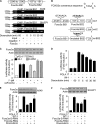
Foxo3a can negatively regulate miR-21 expression. A, Foxo3a RNAi induces a reduction of Foxo3a expression levels. A549 cells were infected with the RNAi constructs of Foxo3a or its scramble form (Foxo3a-S-RNAi). Cells were harvested at the indicated times for the analysis of Foxo3a and P-Foxo3a by immunoblotting. B and C, knockdown of Foxo3a attenuates the reduction of miR-21 levels induced by doxorubicin. A549 (B) and SH-EP1 cells (C) were infected with the RNAi constructs of Foxo3a or its scramble form (Foxo3a-S-RNAi). 24 h after infection cells were treated with 2 μm doxorubicin. 1 h after doxorubicin treatment miR-21 was analyzed by qRT-PCR. The levels of miR-21 analyzed by qRT-PCR were normalized to that of U6. *, p < 0.05 versus doxorubicin alone. D, enforced expression of the constitutively active form of Foxo3a (caFoxo3a) is shown. A549 cells were infected with the adenoviral construct of caFoxo3a. The adenovirus containing β-galactosidase (β-gal) was used as a control. Cells were harvested at the indicated times for the analysis of caFoxo3a expression by immunoblot. E and F, enforced expression of caFoxo3a leads to a reduction of miR-21 levels. A549 (E) and SH-EP1 cells (F) were infected with the adenoviral construct of caFoxo3a. The adenovirus containing β-galactosidase was used as a control. Cells were harvested at the indicated times for the analysis of miR-21 expression by qRT-PCR. The levels of miR-21 analyzed by qRT-PCR were normalized to that of U6. *, p < 0.05 versus control. Data are expressed as the means ± S.E. of three independent experiments.
FIGURE 3.
FIGURE 3.
Foxa3a and miR-21 cross-talk in apoptosis. A and B, enforced expression of miR-21 attenuates apoptosis induced by caFoxo3a. A549 (A) or SH-EP1 cells (B) were co-infected with the adenoviral construct of caFoxo3a or miR-21. The adenovirus containing β-galactosidase (β-gal) was used as a control. Apoptosis was analyzed by TUNEL assay 48 h after infection. TUNEL-positive cells were counted under the fluorescent microscope according to the kit instructions. *, p < 0.05 versus caFoxo3a alone. C, enforced expression of miR-21 inhibits apoptosis induced by doxorubicin. A549 cells were infected with the adenoviral construct of miR-21 or β-gal. Cells were treated with 2 μm doxorubicin 24 h after infection. A TUNEL assay was performed 12 h after doxorubicin treatment. The percentages of TUNEL-positive cells are shown in the left panel. *, p < 0.05 versus doxorubicin alone. Representative photos of TUNEL staining are shown in the right panel. Bar = 50 μm. The nuclei with the green color are TUNEL-positive. The nuclei were counterstained with 4′,6-diamidino-2-phenylindole (DAPI, blue). D, knockdown of miR-21 sensitizes doxorubicin to inducing apoptosis is shown. A549 cells were transfected with miR-21 antagomir or the antagomir-NC. 24 h after transfection cells were treated with 0.2 μm doxorubicin. A TUNEL assay was performed 12 h after doxorubicin treatment. *, p < 0.05 versus doxorubicin alone or antagomir alone. E, knockdown of endogenous Foxo3a attenuates apoptosis induced by doxorubicin is shown. A549 cells were infected with the RNAi constructs of Foxo3a or its scramble form (Foxo3a-S-RNAi). Cells were then transfected with miR-21 antagomir or the antagomir-NC. 24 h after transfection cells were treated with 2 μm doxorubicin. Apoptosis was analyzed by TUNEL assay. *, p < 0.05 versus doxorubicin alone. Data are expressed as the mean ± S.E. of three independent experiments.
FIGURE 4.
FIGURE 4.
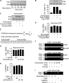
miR-21 is a transcriptional target of Foxo3a. A and B, Foxo3a binds to the miR-21 promoter. The promoter region of miR-21 contains two optimal Foxo3a binding sites. Cells were treated with 2 μm doxorubicin and harvested at the indicated times for the ChIP analysis. Chromatin-bound DNA was immunoprecipitated with the anti-Foxo3a antibody. Anti-FasL antibody was used as a negative control (A). The wild type miR-21 promoters (wt-1 and wt-2) were linked to luciferase (Luc) reporter gene. The mutations were introduced to the consensus binding site-2 (m-BS2) (B). C, Foxo3a inhibits miR-21 promoter activity. HEK293 cells were infected with adenoviral caFoxo3a or (β-galactosidase (β-gal)). 24 h after infection cells were transfected with the constructs of the empty vector (pGL-4.17), wt-1, wt-2, or the mutated promoter (m-BS2), respectively. The expression levels of Foxo3a were detected by immunoblotting (upper panel). Firefly luciferase activities are shown in the lower panel. D, doxorubicin induces a reduction of miR-21 promoter activity. A549 cells were transfected with the constructs of the empty vector (pGL-4.17) or miR-21 wt-1. 24 h after transfection cells were treated with 2 μm doxorubicin and collected at the indicated times for luciferase assay. E and F, knockdown of Foxo3a attenuates the reduction of the promoter activity upon treatment with doxorubicin. A549 (E) and SH-EP1 cells (F) were infected with the adenoviral RNAi constructs of Foxo3a or its scramble form (Foxo3a-S-RNAi) and then transfected with the constructs of the empty vector (pGL-4.17) or miR-21 wt-1 promoter. Cells were treated with 2 μm doxorubicin. Firefly luciferase activities are shown in the lower panel. The expression levels of Foxo3a were detected by immunoblot (upper panel). *, p < 0.05 versus wt-1 plus doxorubicin. Data are expressed as the mean ± S.E. of three independent experiments.
FIGURE 5.
FIGURE 5.
Foxo3a regulates FasL expression independent of the transcriptional manner. A, doxorubicin stimulates FasL expression. A549 cells were treated with 2 μm doxorubicin and harvested at the indicated times for the analysis of FasL by immunoblotting. B, knockdown of FasL attenuates cell death induced by doxorubicin. A549 cells were infected with the adenoviral RNAi constructs of FasL or its scramble form (FasL-S-RNAi) and then treated with 2 μm doxorubicin. FasL was detected by immunoblot. Cell death was analyzed by trypan blue exclusion 24 h after doxorubicin treatment. *, p < 0.05 versus doxorubicin alone. C and D, knockdown of Foxo3a reduces FasL protein but not mRNA expression levels. A549 cells were infected with the adenoviral RNAi constructs of Foxo3a or its scramble form (Foxo3a-S-RNAi) and then treated with 2 μm doxorubicin. FasL protein levels were analyzed by immunoblotting (C), and its mRNA levels were detected by qRT-PCR (D). The values of FasL mRNA analyzed by qRT-PCR were normalized to that of glyceraldehyde-3-phosphate dehydrogenase. E, the promoter region of human FasL contains two Foxo3a consensus binding sites. F, Foxo3a does not bind to FasL promoter, analyzed by ChIP assay. Cells were treated with 2 μm doxorubicin and harvested at the indicated times for the ChIP assay. PCR primers encompassing Foxo3a potential binding site-1 (BS1) and binding site-2 (BS2) were used. G, caFoxo3a induces no significant alterations in FasL mRNA levels. A549 cells were infected with the adenoviral caFoxo3a. The adenoviral β-galactosidase (β-gal) was used as a control. Cells were harvested at the indicated times for the analysis of FasL mRNA by qRT-PCR. The values of FasL mRNA analyzed by qRT-PCR were normalized to that of glyceraldehyde-3-phosphate dehydrogenase. H, caFoxo3a induces no significant changes in FasL promoter activity. A549 cells were infected with the adenoviral caFoxo3a or β-galactosidase and then transfected with the luciferase construct of FasL (FasL-Luc) or the empty vector pGL4.17. Firefly luciferase activities were normalized to Renilla luciferase activities. Data are expressed the ratios of FasL-Luc/the empty vector pGL4.17. I, NFAT4 binds to FasL promoter analyzed by ChIP assay. The primary cardiac fibroblasts were treated with 2 μm doxorubicin and harvested at the indicated time for the ChIP assay. The anti-actin antibody was used as a negative control. The results are expressed as the mean ± S.E. of three independent experiments.
FIGURE 6.
FIGURE 6.
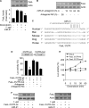
FasL is a target of miR-21. A, enforced expression of miR-21 attenuates FasL elevations induced by doxorubicin. A549 cells were infected with the adenoviral miR-21 or β-gal. 24 h after infection cells were treated with 2 μm doxorubicin. Cells were harvested 1 h after doxorubicin treatment for FasL analysis by immunoblotting. The upper panel shows a representative blot. The lower panel shows the quantitative analysis of FasL levels. The films were densitometrically scanned using NIH ImageJ. The ratios of FasL to actin are shown. *, p < 0.05 versus doxorubicin alone. Data are expressed as the mean ± S.E. of three independent experiments. β-gal, β-galactosidase. B, knockdown of miR-21 leads to an elevation in FasL levels. A549 cells were transfected with miR-21 antagomir or the antagomir-NC. Cells were harvested at the indicate times for FasL analysis by immunoblotting. C, the miR-21 targeting sites in FasL 3′-UTR are conserved. D, miR-21 suppresses FasL translation. HEK293 cells were transfected with the luciferase constructs of wild type FasL-3′-UTR (FasL-3′-UTR-wt) or the mutated FasL-3′-UTR (FasL-3′-UTR-mut) along with the expression plasmids for miR-21, miR-21 antagomir or antagomir-NC. *, p < 0.05 versus FasL-3′-UTR-wt. E, doxorubicin can promote the translational activity of wild type FasL-3′-UTR but not the mutated FasL-3′-UTR (FasL-3′-UTR-mut). A549 cells were transfected with the luciferase constructs of wild type FasL-3′-UTR or the FasL-3′-UTR-mut and then treated with 2 μm doxorubicin and harvested at the indicated times for the luciferase assay. The luciferase activity (Luc) of each construct before doxorubicin treatment was taken as 100%. F, miR-21 can suppress the expression of FasL with wild type but not mutated 3′-UTR. A549 cells were co-infected with the adenoviral miR-21, β-galactosidase, wild type FasL-3′-UTR (FasL-3′-UTR-wt) or the mutated FasL-3′-UTR (FasL-3′-UTR-mut). FasL expression was detected by immunoblotting.

Similar articles

Cited by

References

    1. Park S. M., Schickel R., Peter M. E. (2005) Curr. Opin. Cell Biol. 17, 610–616 - PubMed
    1. He L., He X., Lim L. P., de Stanchina E., Xuan Z., Liang Y., Xue W., Zender L., Magnus J., Ridzon D., Jackson A. L., Linsley P. S., Chen C., Lowe S. W., Cleary M. A., Hannon G. J. (2007) Nature 447, 1130–1134 - PMC - PubMed
    1. Nasser M. W., Datta J., Nuovo G., Kutay H., Motiwala T., Majumder S., Wang B., Suster S., Jacob S. T., Ghoshal K. (2008) J. Biol. Chem. 283, 33394–33405 - PMC - PubMed
    1. Löffler D., Brocke-Heidrich K., Pfeifer G., Stocsits C., Hackermüller J., Kretzschmar A. K., Burger R., Gramatzki M., Blumert C., Bauer K., Cvijic H., Ullmann A. K., Stadler P. F., Horn F. (2007) Blood 110, 1330–1333 - PubMed
    1. Accili D., Arden K. C. (2004) Cell 117, 421–426 - PubMed

Publication types