. 2010 Sep;24(9):3117-21.
doi: 10.1096/fj.09-151381. Epub 2010 Apr 6.

Beyond 9+0: noncanonical axoneme structures characterize sensory cilia from protists to humans

Affiliations

Beyond 9+0: noncanonical axoneme structures characterize sensory cilia from protists to humans

Eva Gluenz et al. FASEB J. 2010 Sep.

Abstract

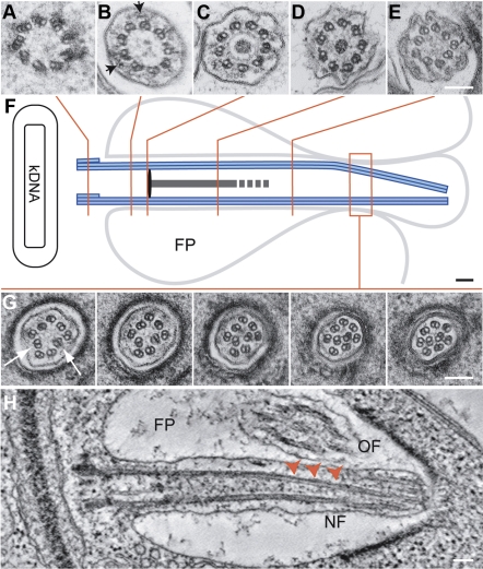
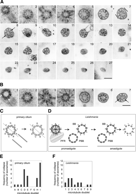
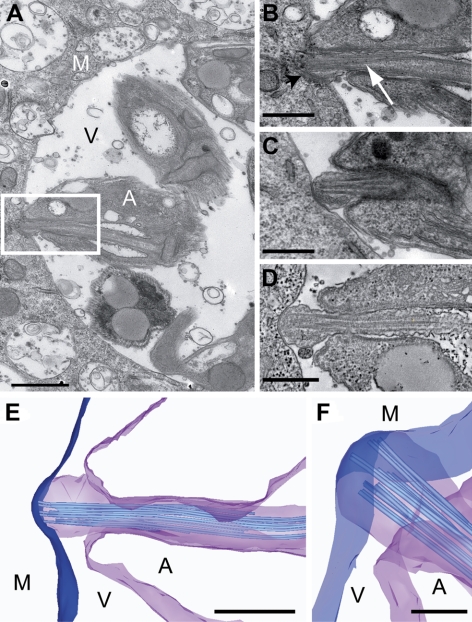
The intracellular amastigote stages of parasites such as Leishmania are often referred to as aflagellate. They do, however, possess a short axoneme of cryptic function. Here, our examination of the structure of this axoneme leads to a testable hypothesis of its role in the cell biology of pathogenicity. We show a striking similarity between the microtubule axoneme structure of the Leishmania mexicana parasite infecting a macrophage and vertebrate primary cilia. In both, the 9-fold microtubule doublet symmetry is broken by the incursion of one or more microtubule doublets into the axoneme core, giving rise to an architecture that we term here the 9v (variable) axoneme. Three-dimensional reconstructions revealed that no particular doublet initiated the symmetry break, and moreover it often involved 2 doublets. The tip of the L. mexicana flagellum was frequently intimately associated with the macrophage vacuole membrane. We propose that the main function of the amastigote flagellum is to act as a sensory organelle with important functions in host-parasite interactions and signaling in the intracellular stage of the L. mexicana life cycle.


Figures

Figure 1.
Figure 1.
Structure of the L. mexicana amastigote flagellum. J774 macrophages were infected with stationary phase L. mexicana promastigotes . Three days postinfection cells were fixed and processed for TEM essentially as described previously . A–E, G) Arrangement of microtubules along the axoneme. Arrowheads (B) indicate IFT particles. F) Position of sections in A–E and G. G) Displacement of doublets (arrow) shown in serial thin sections through the distal portion. H) Tomographic slice from a reconstructed 3-D volume of a dividing amastigote showing the growing flagellum (NF) in longitudinal section; arrowheads indicate symmetry break. FP, flagellar pocket; OF, mature flagellum. Scale bars = 100 nm.
Figure 2.
Figure 2.
Displacement of doublet microtubules in kidney primary cilia and L. mexicana flagella. A, B) IMCD3 cells were processed for TEM as described previously . Numbers indicate adjacent serial sections along the IMCD3 primary cilium. Proximally, the axoneme is 9 + 0 (A). Flattening (A7, lines) is the first sign of doublet displacement (A8, B7; arrows). The doublet-membrane connections remain for much of the cilium length (e.g., A16, inset, arrow). C–F) mapping doublet displacement in primary cilia (C, E) and L. mexicana (D, F). C) The basal body-procentriole pair with letters (A–I) assigned to each triplet relative to procentriole position; these positions were followed (arrow) through each series. D) Absolute doublet and triplet numbering show that the L. mexicana promastigotes probasal body (PBB) is always adjacent to basal-body (BB) triplet 7. These numbers were translated onto the amastigote BB/PBB pair (dashed arrow) and their positions followed as in C. E, F) The frequency of doublet displacement observed for each doublet in 18 primary cilium axonemes (E; 2 doublets were displaced simultaneously in 14 axonemes) and in 11 amastigote axonemes (F; 2 doublets were displaced simultaneously in 6 axonemes). Scale bars = 200 nm.
Figure 3.
Figure 3.
The amastigote flagellum is in intimate contact with the parasitophorous vacuole membrane. A) Thin section TEM view of L. mexicana amastigotes in a J774 macrophage vacuole. B) Higher-magnification view of the area delineated by the white box in A. Amastigote flagellum tip is closely associated with the vacuole membrane (black arrowhead). White arrow indicates doublet displacement. C) Further example of the flagellum-vacuole membrane junction. D) Tomographic slice from a reconstructed 3-D volume of an amastigote flagellum in close contact with the vacuole membrane. E, F) Tomography model views (23) of the flagellum-vacuole membrane junction. A, L. mexicana amastigote; M, macrophage; V, vacuole. Scale bars = 500 nm (A–E); 200 nm (F).

References

    1. Gerdes J M, Davis E E, Katsanis N. The vertebrate primary cilium in development, homeostasis, and disease. Cell. 2009;137:32–45. - PMC - PubMed
    1. Gull K. The parasite point of view: insights and questions on the cell biology of Trypanosoma and Leishmania parasite-phagocyte interactions. Russell D G, Gordon S, editors. Washington, DC: ASM Press; Phagocyte-Pathogen Interactions: Macrophages and the Host Response to Infection. 2009:453–462.
    1. Simpson C F, Harvey J W, French T W. Ultrastructure of amastigotes of Leishmania donovani in the bone marrow of a dog. Am J Vet Res. 1982;43:1684–1686. - PubMed
    1. Alexander J. Unusual axonemal doublet arrangements in the flagellum of Leishmania amastigotes. Trans R Soc Trop Med Hyg. 1978;72:345–347. - PubMed
    1. Hentzer B, Kobayasi T. The ultrastructure of Leishmania tropica in skin lesions. Acta Pathol Microbiol Scand B. 1977;85:153–160. - PubMed

Publication types

LinkOut - more resources