. 2010 Aug;24(8):2859-68.
doi: 10.1096/fj.09-148007. Epub 2010 Apr 6.

Primary cilium-dependent mechanosensing is mediated by adenylyl cyclase 6 and cyclic AMP in bone cells

Affiliations

Primary cilium-dependent mechanosensing is mediated by adenylyl cyclase 6 and cyclic AMP in bone cells

Ronald Y Kwon et al. FASEB J. 2010 Aug.

Abstract

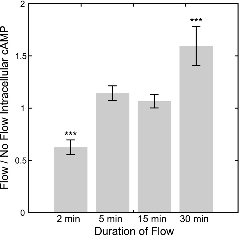
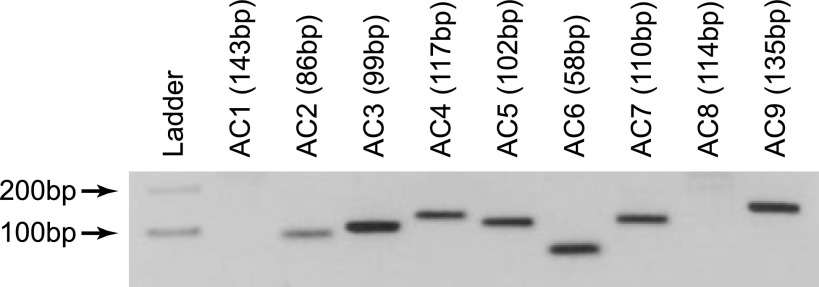
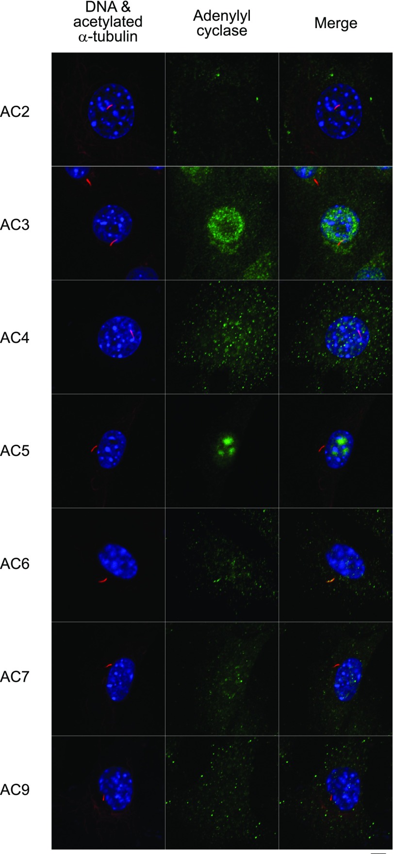
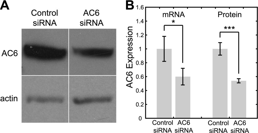
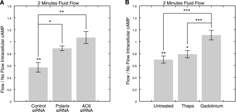
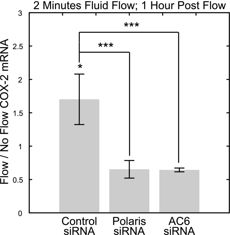
Primary cilia are chemosensing and mechanosensing organelles that regulate remarkably diverse processes in a variety of cells. We previously showed that primary cilia play a role in mediating mechanosensing in bone cells through an unknown mechanism that does not involve extracellular Ca(2+)-dependent intracellular Ca(2+) release, which has been implicated in all other cells that transduce mechanical signals via the cilium. Here, we identify a molecular mechanism linking primary cilia and bone cell mechanotransduction that involves adenylyl cyclase 6 (AC6) and cAMP. Intracellular cAMP was quantified in MLO-Y4 cells exposed to dynamic flow, and AC6 and primary cilia were inhibited using RNA interference. When exposed to flow, cells rapidly (<2 min) and transiently decreased cAMP production in a primary cilium-dependent manner. RT-PCR revealed differential expression of the membrane-bound isoforms of adenylyl cyclase, while immunostaining revealed one, AC6, preferentially localized to the cilium. Further studies showed that decreases in cAMP in response to flow were dependent on AC6 and Gd(3+)-sensitive channels but not intracellular Ca(2+) release and that this response mediated flow-induced COX-2 gene expression. The signaling events identified provide important details of a novel early mechanosensing mechanism in bone and advances our understanding of how signal transduction occurs at the primary cilium.


Figures

Figure 1
Figure 1
Flow/no flow intracellular cAMP levels in MLO-Y4 osteocyte-like cells immediately following 2, 5, 15, and 30 min of flow exposure. Flow/no flow ratio <1 or >1 indicates a decrease or increase of cAMP in response to flow, respectively. Oscillatory flow induced a rapid and transient decrease in cAMP levels after 2 min of exposure and an increase after 30 min of exposure. ***P < 0.001.
Figure 2
Figure 2
RT-PCR targeting mouse AC1–9 reveals MLO-Y4 cells express AC2–7 and AC9.
Figure 3
Figure 3
Immunostaining for primary cilia, identified as linear structures enriched in acetylated α-tubulin, (red) and adenylyl cyclase (green). Nuclei are counterstained with DAPI (blue). Of the expressed isoforms (AC2–7 and AC9), only AC6 was found to preferentially localize to the primary cilium. Scale bar = 5 μm.
Figure 4
Figure 4
Treatment with AC6 siRNA results in significant decreases in AC6 expression at both gene and protein levels. A) Western blot of AC6 in cells treated with control and AC6 siRNA. Samples are normalized by actin. B) AC6 gene expression, as quantified by real time RT-PCR, and protein expression, as quantified by Western blot densitometry, in cells treated with control and AC6 siRNA. *P < 0.05, ***P < 0.001.
Figure 5
Figure 5
Polaris and AC6 depletion inhibits flow-induced decreases in cAMP via a mechanism that involves Gd3+-sensitive ion channels but not release of Ca2+ from intracellular stores. A) Flow/no flow intracellular cAMP levels immediately following 2 min of flow exposure in cells treated with control, polaris, and AC6 siRNA. Flow-induced decreases in intracellular cAMP levels were significantly inhibited in polaris- and AC6-depleted cells. B) Flow/no flow intracellular cAMP levels following 2 min of flow in untreated cells and cells treated with thapsigargin or Gd3+. Intracellular cAMP levels significantly decreased in response to flow in untreated cells and cells treated with thapsigargin; however this response was lost in cells treated with Gd3+. *P < 0.05, **P < 0.01, ***P < 0.001.
Figure 6
Figure 6
Two minutes of fluid flow induces COX-2 gene expression in a primary cilium- and AC6-dependent manner. Flow/no flow COX-2 mRNA levels following 2 min of flow exposure and a 1-h postflow incubation period indicate that COX-2 expression was significantly increased in response to flow exposure in cells treated with control siRNA but not in cells treated with polaris and AC6 siRNA. *P < 0.05, ***P < 0.001.

Similar articles

Cited by

References

    1. Ingber D. E. Mechanobiology and diseases of mechanotransduction. Ann Med. 2003;35:1–14. - PubMed
    1. Knothe Tate M. L. “Whither flows the fluid in bone?” An osteocyte’s perspective. J Biomech. 2003;36:1409–1424. - PubMed
    1. Malone A. M., Anderson C. T., Tummala P., Kwon R. Y., Johnston T. R., Stearns T., Jacobs C. R. Primary cilia mediate mechanosensing in bone cells by a calcium-independent pathway. Proc Natl Acad Sci U S A. 2007;104:13325–13330. - PMC - PubMed
    1. Klein-Nulend J., Semeins C. M., Ajubi N. E., Nijweide P. J., Burger E. H. Pulsating fluid flow increases nitric oxide (NO) synthesis by osteocytes but not periosteal fibroblasts—correlation with prostaglandin upregulation. Biochem Biophys Res Commun. 1995;217:640–648. - PubMed
    1. Vezeridis P. S., Semeins C. M., Chen Q., Klein-Nulend J. Osteocytes subjected to pulsating fluid flow regulate osteoblast proliferation and differentiation. Biochem Biophys Res Commun. 2006;348:1082–1088. - PubMed

Publication types