. 2010 Jun;24(6):1136-50.
doi: 10.1210/me.2009-0466. Epub 2010 Apr 6.

Glycogen synthase kinase-3 plays a central role in mediating glucocorticoid-induced apoptosis

Affiliations

Glycogen synthase kinase-3 plays a central role in mediating glucocorticoid-induced apoptosis

Rachel Spokoini et al. Mol Endocrinol. 2010 Jun.

Abstract

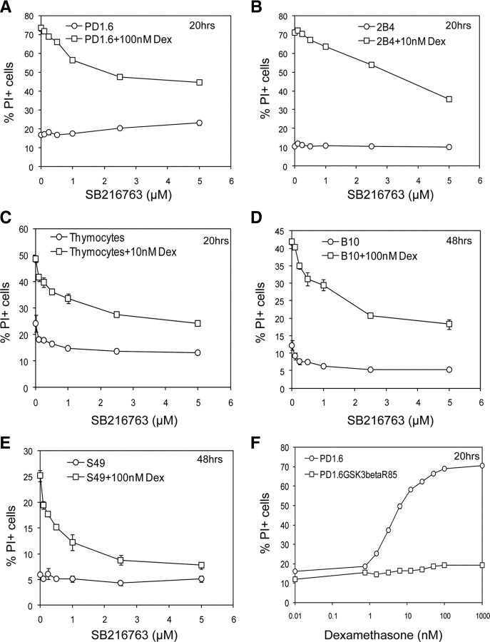
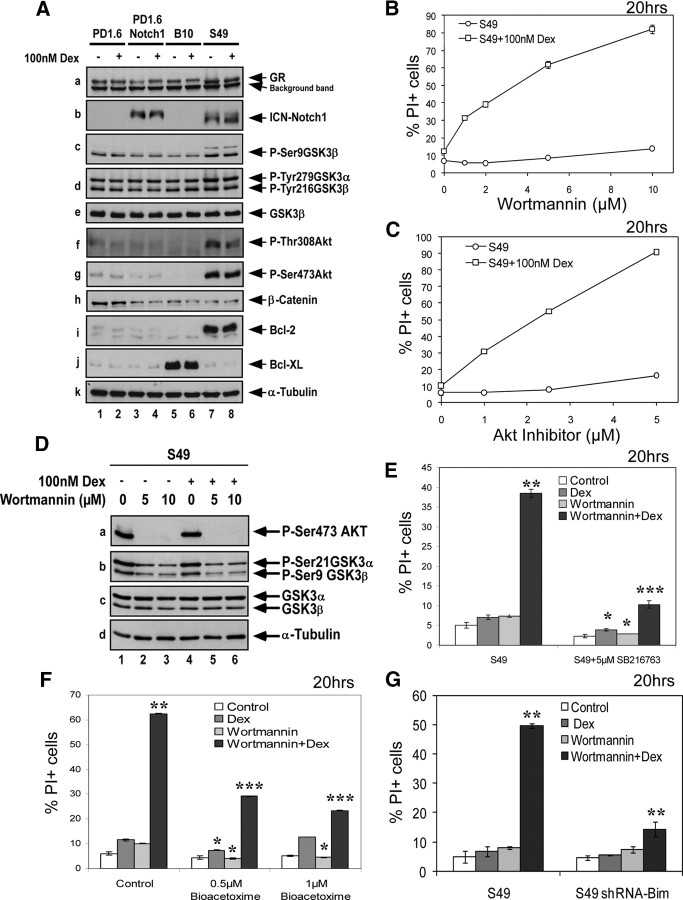
It is still unclear how glucocorticoids (GCs) induce apoptosis of thymocytes and T lymphoma cells. Emergence of GC-resistant lymphoma cells is a major obstacle in GC therapy, emphasizing the need for novel strategies that maintain the sensitivity of lymphoma cells to the proapoptotic effects of GC. We have undertaken a kinome study to elucidate the signal transduction pathways involved in mediating GC-induced apoptosis. Our study shows that glycogen synthase kinase (GSK3) plays a central role in promoting GC-induced apoptosis. In the absence of a ligand, GSK3alpha, but not GSK3beta, is sequestered to the glucocorticoid receptor (GR). Exposure to GCs leads to dissociation of GSK3alpha from GR and subsequent interaction of GSK3alpha and GSK3beta with the proapoptotic Bim protein, an essential mediator of GC-induced apoptosis. Chemical inhibition of GSK3 by SB216763, BIO-Acetoxime, or LiCl and GSK3 inhibition using a dominant-negative mutant of GSK3 impede this cell death process, indicating that GSK3 is involved in transmitting the apoptotic signal. GC resistance in lymphoma cells can be relieved by inhibiting the phosphatidylinositol-3 kinase-Akt survival pathway, which inactivates GSK3. Notch1, a transcription factor frequently activated in T acute lymphoblastic leukemia cells, confers GC resistance through activation of Akt. Altogether, this study illuminates the link connecting upstream GR signals to the downstream mediators of GC-induced apoptosis. Our data suggest that targeting protein kinases involved in GSK3 inactivation should improve the outcome of GC therapy.


Figures

Fig. 1.
Fig. 1.
Effect of protein kinase inhibitors on GC-induced apoptosis. Thymocytes (A), PD1.6 T lymphoma (B), S49 T lymphoma (C), and CCRF-CEM T ALL (D) cells were incubated with the indicated concentrations of Dex in the absence or presence of various protein kinase inhibitors for 20 h (A–C), 48 h (C and D), or 72 h (D). Apoptosis was measured by the PI-exclusion assay, which is the most adequate assay for assessing cell death in lymphoma and leukemia cells (32 ). The results of the PI-exclusion assay correlate with the caspase-3 activation assay. PP1, a Src inhibitor; SB203580, a p38 inhibitor; PD98059, a MEK inhibitor; SP600125, a JNK inhibitor; roscovitine, a Cdk2 inhibitor; SB216763, a GSK3 inhibitor. The casein kinase I (CKI) inhibitor was obtained from Calbiochem and the casein kinase II (CKII) inhibitor is 4,5,6,7-tetrabromobenzotriazole. *, P < 0.05; **, P < 0.005; ***, P < 0.0005. Cont, Control.
Fig. 1.
Fig. 1.
Effect of protein kinase inhibitors on GC-induced apoptosis. Thymocytes (A), PD1.6 T lymphoma (B), S49 T lymphoma (C), and CCRF-CEM T ALL (D) cells were incubated with the indicated concentrations of Dex in the absence or presence of various protein kinase inhibitors for 20 h (A–C), 48 h (C and D), or 72 h (D). Apoptosis was measured by the PI-exclusion assay, which is the most adequate assay for assessing cell death in lymphoma and leukemia cells (32 ). The results of the PI-exclusion assay correlate with the caspase-3 activation assay. PP1, a Src inhibitor; SB203580, a p38 inhibitor; PD98059, a MEK inhibitor; SP600125, a JNK inhibitor; roscovitine, a Cdk2 inhibitor; SB216763, a GSK3 inhibitor. The casein kinase I (CKI) inhibitor was obtained from Calbiochem and the casein kinase II (CKII) inhibitor is 4,5,6,7-tetrabromobenzotriazole. *, P < 0.05; **, P < 0.005; ***, P < 0.0005. Cont, Control.
Fig. 2.
Fig. 2.
Dex-induced apoptosis is prevented by inhibition of GSK3. A–E, The specific GSK3 inhibitor SB216763 prevents Dex-induced apoptosis of T lymphoma cells and thymocytes. The highly GC-sensitive PD1.6 T lymphoma (A), 2B4 T hybridoma (B), and thymocytes (C), as well as the partially resistant B10 (D) and S49 (E) T lymphoma cells, were treated with various concentrations of the GSK3 inhibitor SB216763 in the presence or absence of Dex for 20 h (A–C) or 48 h (D and E). Apoptosis was measured by the PI-exclusion assay. F, PD1.6 expressing a kinase-inactive variant of GSK3 (PD1.6GSK3βR85) becomes resistant to Dex-induced apoptosis. The cells were treated for 20 h with different Dex concentrations, and apoptosis was determined by PI exclusion. P < 0.001.
Fig. 3.
Fig. 3.
GSK3α interacts with GR. A, The effect of Dex treatment on GSK3 phosphorylation. PD1.6 cells were untreated or treated with 100 nm Dex for the indicated time intervals and processed for Western blotting (WB) using antibodies to phospho-Ser9 GSK3β, phospho-Tyr216 GSK3β, GSK3β, or α-Tubulin. B, GSK3α interacts with native GR and is released upon Dex treatment. Thymocytes were untreated or treated with 100 nm Dex for the indicated time intervals. Total lysates were immunoprecipitated (IP) using M20 antibodies specific to GR (Santa Cruz Biotechnology) and Protein A beads. The one-step Genscript IP-WB kit was used for Western blot analysis to prevent the detection of heavy chain. Nevertheless, a band of the heavy chain is seen, as indicated. Antibodies to GSK3α/β (0011-Α, Santa Cruz Biotechnology) were applied. For the IP analysis, two pre-IP samples (lanes 1 and 9; input 1:40 of IP samples) were run to display the location of GSK3α and -β. Lane 2 contains only the antibody (Ab), which provides the location of the heavy chain. Samples of total lysates were taken before immunoprecipitation (Pre-IP), to show the initial expression levels of the indicated proteins. C, GR interacts with GSK3α in PD1.6 T lymphoma cells. PD1.6 cells were untreated (lane 1), or treated with SB216763 and/or Dex for 1 h. Immunoprecipitation was performed as described in panel B. Panels a and b are two different exposures. The GSK3α band appears just above the heavy chain band. D, Selective binding of GSK3α to GR. MCF-7 cells were transfected with 5 μg of plasmids encoding hGRα (lanes 1–3), hGSK3α (lanes 2 and 4), and/or hGSK3β.HA (hemagglutinin-labeled) (lanes 3 and 5). Empty pcDNA3 vector (5 μg) was included in samples 1, 4, and 5 to achieve equal amounts of transfected DNA. Cell lysates were immunoprecipitated with M20 antibodies against GR, and the presence of coimmunoprecipitated GSK3 was analyzed by WB as described in panel B. E, Long-term inhibition of GSK3 leads to reduced GR expression. PD1.6 cells were treated with SB216763 for 2 and 24 h, and the GR expression level was analyzed on Western blot. β-Catenin expression is shown as indication for GSK3 inhibition.
Fig. 4.
Fig. 4.
GSK3α and GSK3β interact with Bim after exposure of thymocytes (A) and 2B4 T cells (B) to Dex. Thymocytes (A) and 2B4 T cells (B) were either untreated or treated with 100 nm Dex for the indicated time intervals. Cell lysates were immunoprecipitated with antibodies to Bim and processed for Western blot analysis using nonreduced protein sample buffer. Coimmunoprecipitated GSK3α and GSK3β were detected by Western blotting as described in Fig. 3B.
Fig. 5.
Fig. 5.
GC resistance of S49 cells is overcome by relieving Akt-mediated inhibition of GSK3. A, The partially resistant S49 T lymphoma cells display increased Akt activation and express elevated levels of antiapoptotic Bcl-2 and intracellular cleaved Notch1 (ICN). Western blot analysis of the highly GC-sensitive PD1.6 T lymphoma, PD1.6 cells overexpressing ICN-Notch1 (PD1.6Notch1), and the partially resistant B10 and S49 T lymphoma cells are presented. The antibody used in panel c is specific for phospho-Ser9 GSK3β. It also detects a slower migrating band in S49 cells, which may be phospho-Ser21 GSK3α. B and C, Inhibition of the PI3K-Akt axis by either wortmannin (B) or the Akt inhibitor VIII (C) confers GC sensitivity on S49 cells. S49 cells were treated with 100 nm Dex alone or in the presence of increasing concentrations of either wortmannin (C) or Akt inhibitor VIII (D) for 20 h before the extent of apoptosis was measured. The use of either drug alone had no apoptotic effect on S49 cells, whereas combined treatment induced strong apoptosis. D, The PI3K inhibitor wortmannin prevents Akt and GSK3 phosphorylation. S49 cells were either untreated or treated with wortmannin and/or Dex for 1 h. Western blot analysis was performed with antibodies to phospho-Ser473 Akt, phospho-Ser21/9 GSK3α/β, GSK3α/β, or α-tubulin. E and F, The GSK3 inhibitors SB216763 (E) and BIO-Acetoxime (F) prevent wortmannin sensitization to Dex. S49 cells were exposed to 5 μm wortmannin, 5 μm SB216763, or 0.5–1 μm BIO-Acetoxime, with or without 100 nm Dex for 20 h before apoptosis was measured. G, shRNA to Bim prevents wortmannin sensitization of S49 cells to Dex-induced apoptosis. Control S49 cells or S49 cells transfected with pSilencer-Bim were exposed to 5 μm wortmannin and/or 100 nm Dex, and the extent of apoptosis was measured 20 h later. *, P < 0.05; **, P < 0.01; ***, P < 0.001.
Fig. 6.
Fig. 6.
Notch1 confers GC resistance by activating Akt with subsequent inhibition of GSK3. A and B, Overexpression of ICN-Notch1 confers GC resistance on 2B4, but not on PD1.6 cells. 2B4 (A), 2B4Notch1 cells overexpressing ICN-Notch1 (A), PD1.6 (B) or PD1.6Notch1 cells overexpressing ICN-Notch1 (B) were exposed to Dex for 20 h. Apoptosis was measured by PI exclusion. C, ICN-Notch1 is transcriptionally active in 2B4 cells, but not in PD1.6 cells. RT-PCR analysis was performed on the indicated cell lines for detection of the mRNA of GR, SRG3, a gene product related to GC sensitivity (86 ), and the Notch-regulated gene Deltex1. The housekeeping gene glyceraldehyde-3-phosphate dehydrogenase (GAPDH) was included as a control. D, 2B4Notch1 cells show elevated Akt phosphorylation. Western blot analysis of untreated or Dex-treated (100 nm, 2 h) 2B4 and 2B4Notch1 cells. E, Mcl-1 is induced in 2B4 cells overexpressing Notch1. Total extracts of the indicated cell lines were analyzed by Western blotting.
Fig. 7.
Fig. 7.
A proposed model for GC-induced apoptosis in thymocytes and T lymphoma cells. This model is based on findings described in the present study combined with data published in the literature. In the absence of ligand, GR is sequestered in the cytosol bound to Hsps and immunophilins (no. 1). A proportion of GR also interacts with GSK3α. Upon exposure to GC, GR is released from the heat shock complex, and GSK3α dissociates from GR (no. 2). GR dimerizes and translocates to the nucleus and the mitochondria (no. 3). The nuclear translocation occurs in both sensitive and resistant T lymphoma cells, whereas mitochondrial translocation takes place only in sensitive cells (Ref. 13 ). In the nucleus, GR alters the transcription of multiple genes through transactivation and transrepression (no. 4). Of importance, the essential proapoptotic Bim is up-regulated, a process that requires proper activation of p38 and the transcription factor FoxO3 (Ref. 35 ). Bim is frequently expressed at a high basal level in many hematopoietic cells (Ref. 1 ). GR translocated to the mitochondria delivers signals (no. 5) that may activate GSK3 (no. 6). GSK3α and GSK3β interact with Bim (no. 7). Bim acts upstream to Bax and Bak (no. 7), which execute the apoptotic cascade (no. 8) (Ref. 87 ). GSK3β may also phosphorylate VDAC (no. 7) leading to dissociation of hexokinase II from the mitochondria and facilitation of Bax binding to the mitochondria (Ref. 65 ). Thus, GC-induced apoptosis is effected by a cooperation between nuclear GR (e.g. up-regulation of Bim), mitochondrial GR (dispatching nongenomic signals), and GSK3 (activating the downstream effectors of the intrinsic apoptotic pathway). AP-1, Activator protein 1; NFκB, nuclear factor-κB.

Similar articles

Cited by

References

    1. Sionov RV, Kfir S, Zafrir E, Cohen O, Zilberman Y, Yefenof E2006. Glucocorticoid-induced apoptosis revisited: a novel role for glucocorticoid receptor translocation to the mitochondria. Cell Cycle 5:1017–1026 - PubMed
    1. Sionov RV, Spokoini R, Kfir-Erenfeld S, Cohen O, Yefenof E2008. Mechanisms regulating the susceptibility of hematopoietic malignancies to glucocorticoid-induced apoptosis. Adv Cancer Res 101:127–248 - PubMed
    1. Wang Z, Malone MH, He H, McColl KS, Distelhorst CW2003. Microarray analysis uncovers the induction of the proapoptotic BH3-only protein Bim in multiple models of glucocorticoid-induced apoptosis. J Biol Chem 278:23861–23867 - PubMed
    1. Puthalakath H, Strasser A2002. Keeping killers on a tight leash: transcriptional and post-translational control of the pro-apoptotic activity of BH3-only proteins. Cell Death Differ 9:505–512 - PubMed
    1. Haller J, Mikics E, Makara GB2008. The effects of non-genomic glucocorticoid mechanisms on bodily functions and the central neural system. A critical evaluation of findings. Front Neuroendocrinol 29:273–291 - PubMed

Publication types

MeSH terms